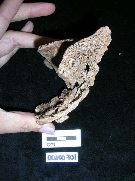
| Site code
|
Context
|
Frame number
|
Photo
|
Description
|
| OCU00
|
20
|
2
|
OCU00_20_2.jpg
|
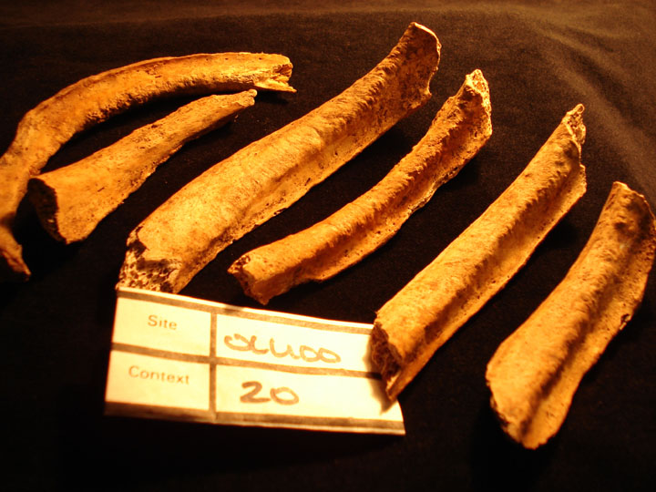
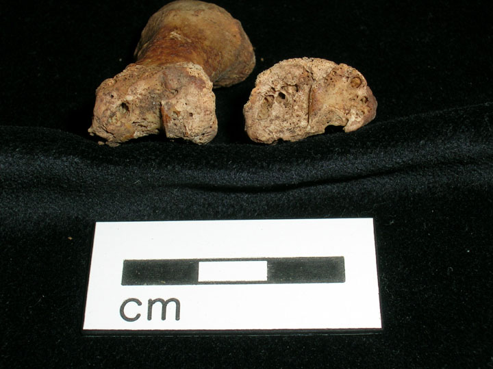
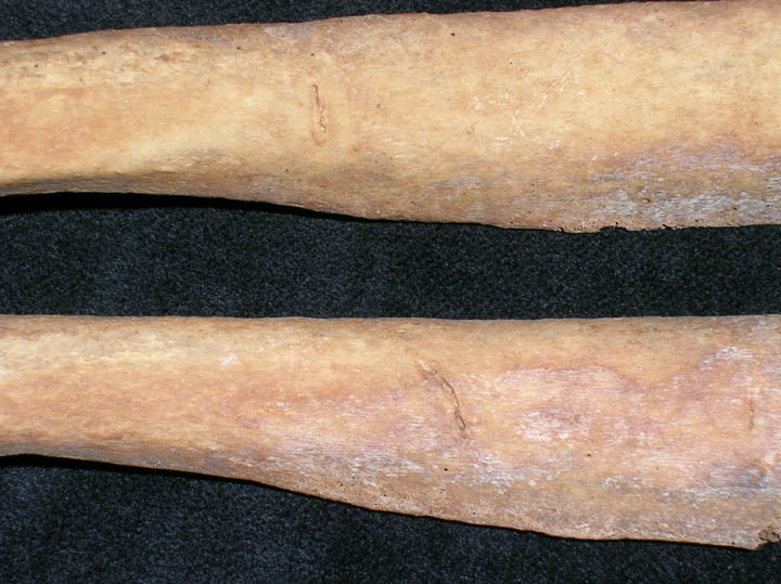
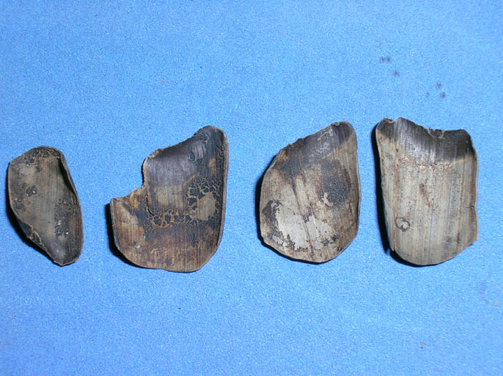
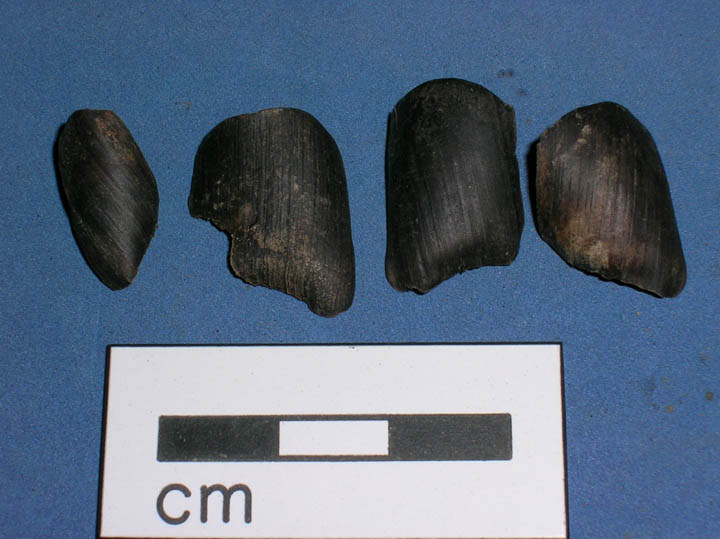
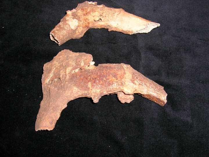
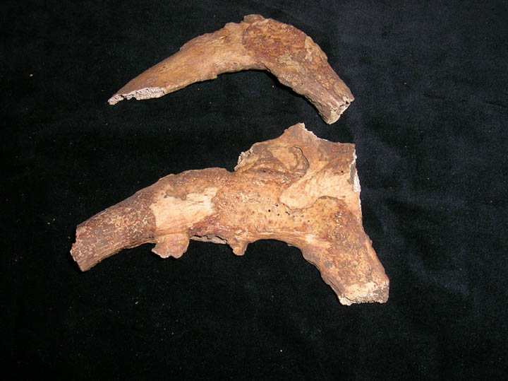
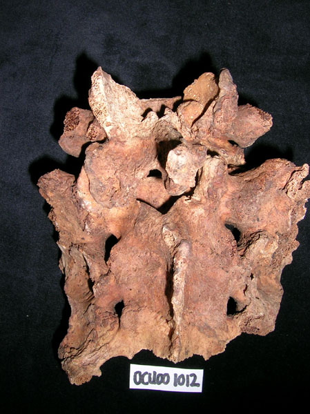
Rib lesions on the visceral surface of several rib shafts, healed and remodelled
|
| OCU00
|
20
|
3
|
OCU00_20_3.jpg
|
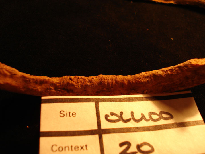
Close up of rib lesions on the visceral surface healed and remodelled
|
| OCU00
|
20
|
4
|
OCU00_20_4.jpg
|
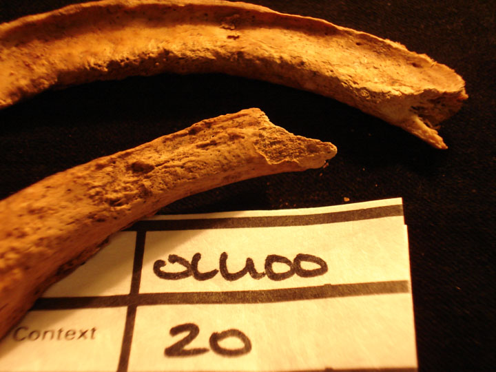
Two rib shafts showing close up of rib lesions on visceral surface healed and remodelled
|
| OCU00
|
35
|
1
|
OCU00_35_1.jpg
|
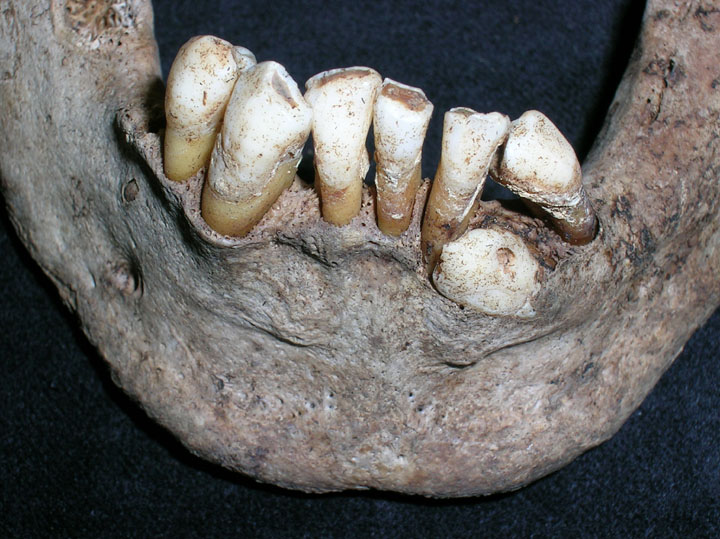
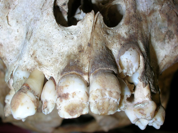
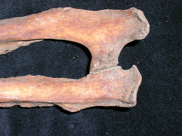
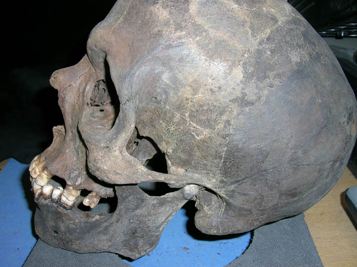
Mandible with impacted canine
|
| OCU00
|
35
|
2
|
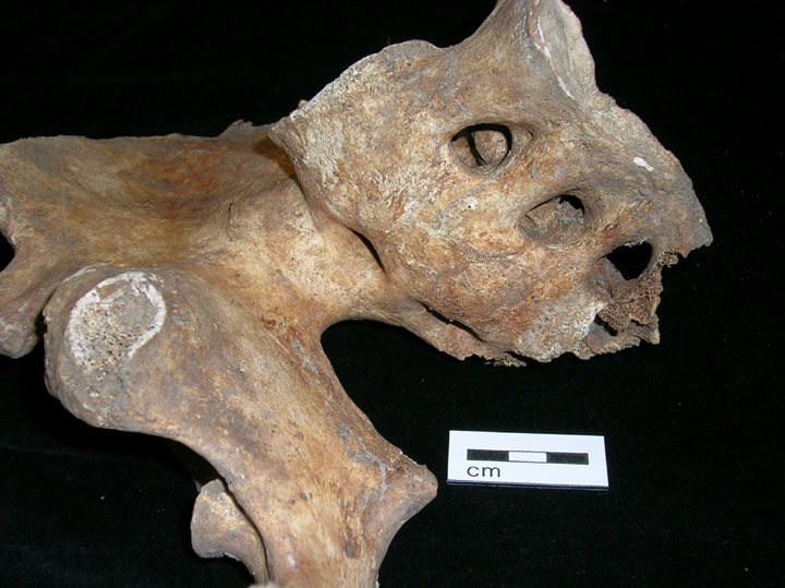
OCU00_35_2.jpg
|
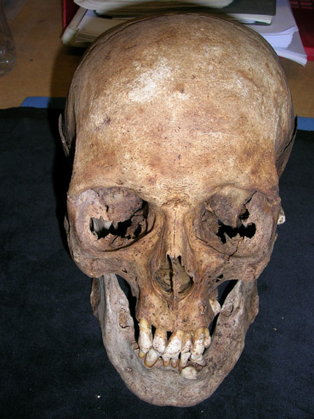
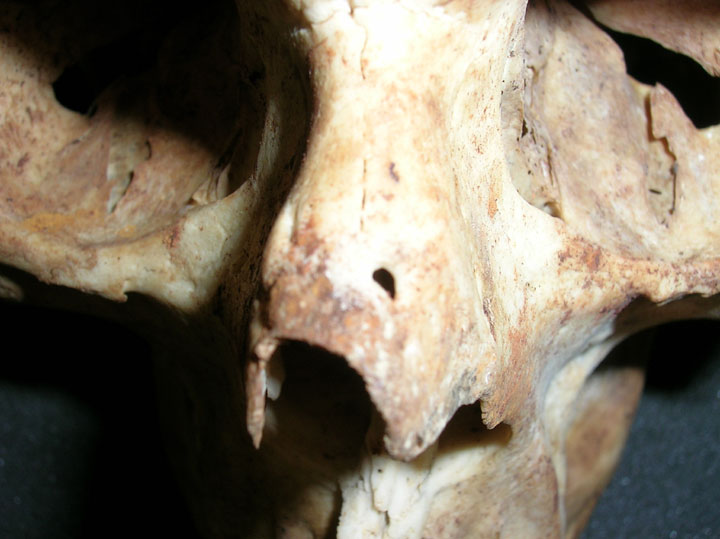
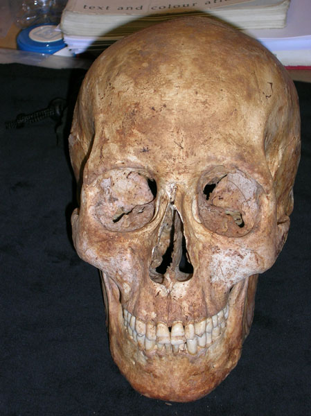
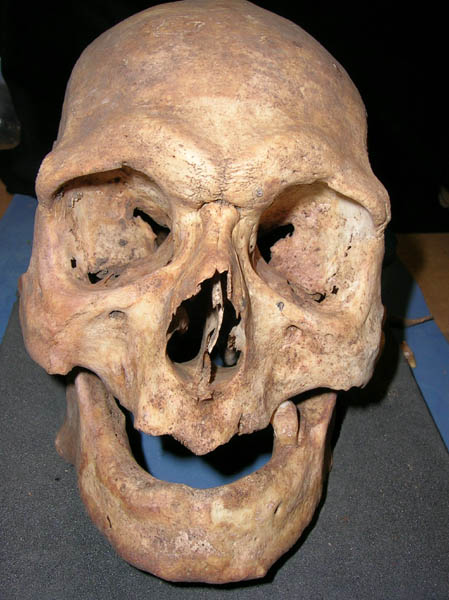
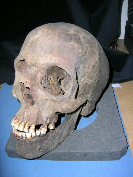
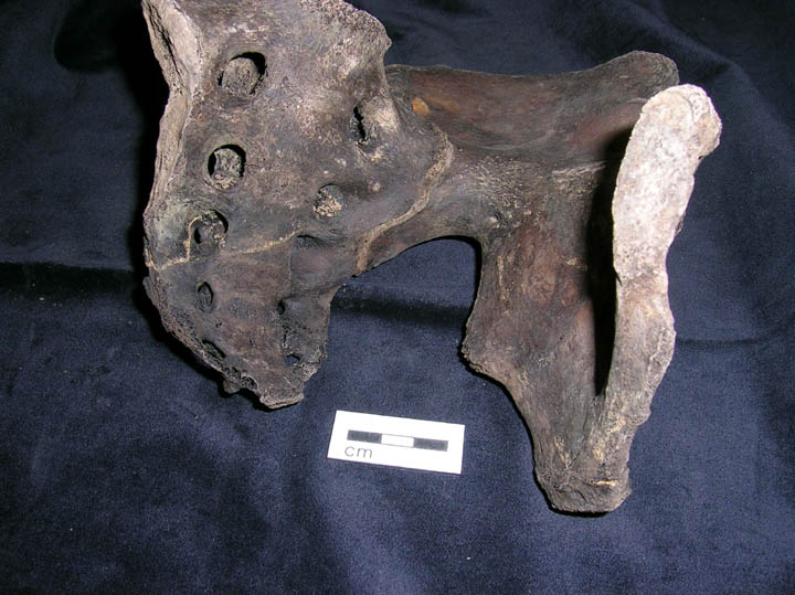
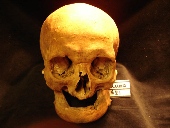
Anterior view of skull
|
| OCU00
|
35
|
3
|
OCU00_35_3.jpg
|
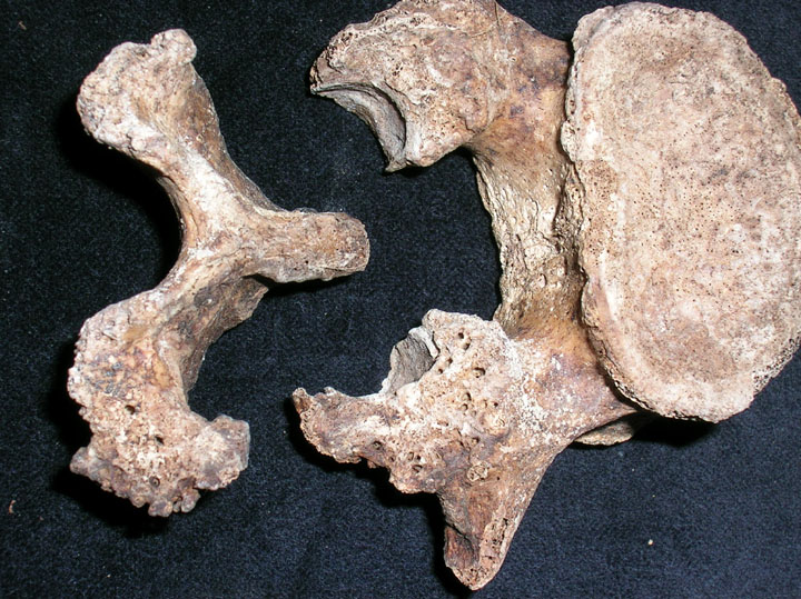
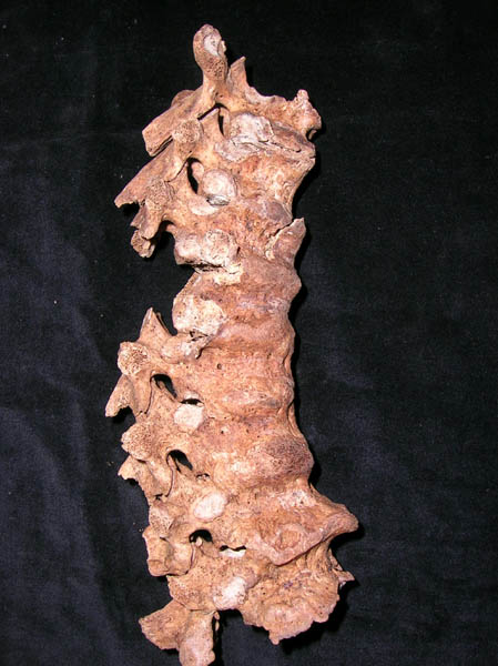
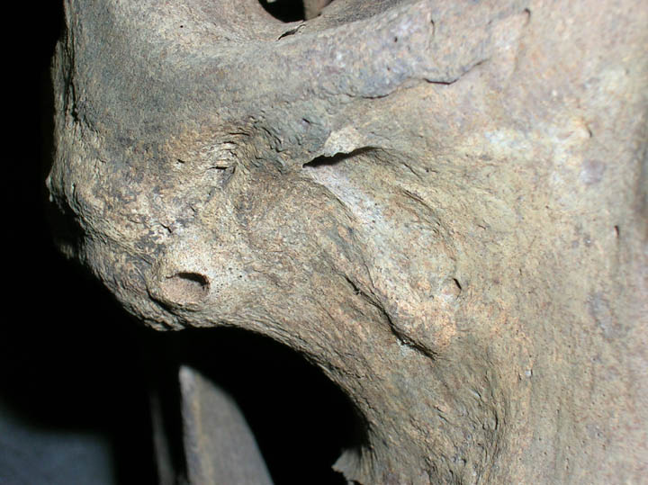
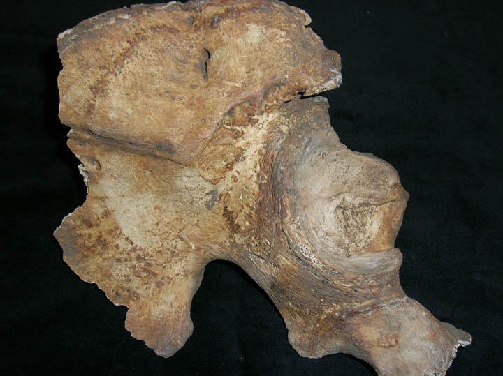
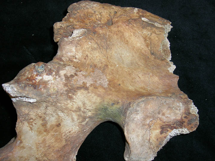
Spondylolisis of 4th Lumbar vertebra
|
| OCU00
|
35
|
4
|
OCU00_35_4.jpg
|
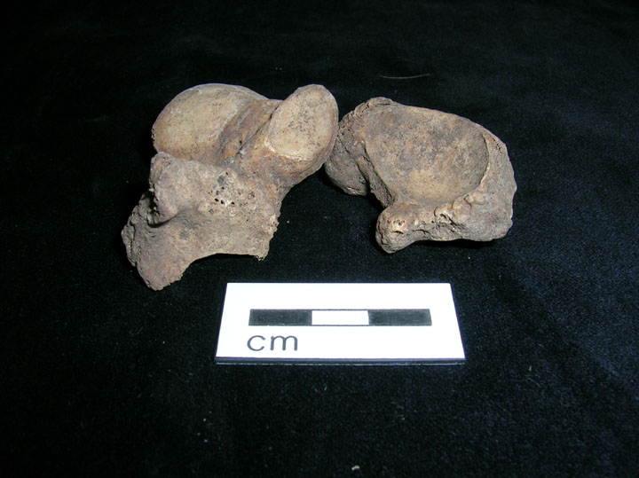
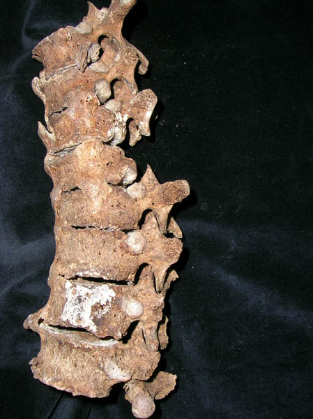
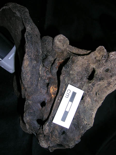
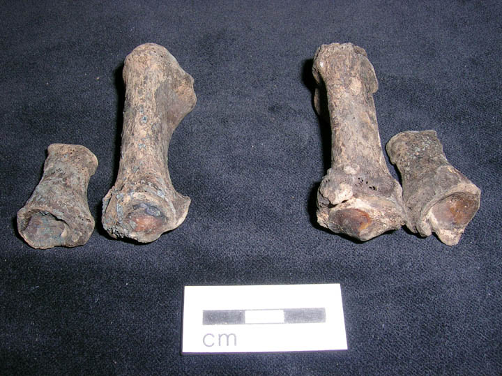
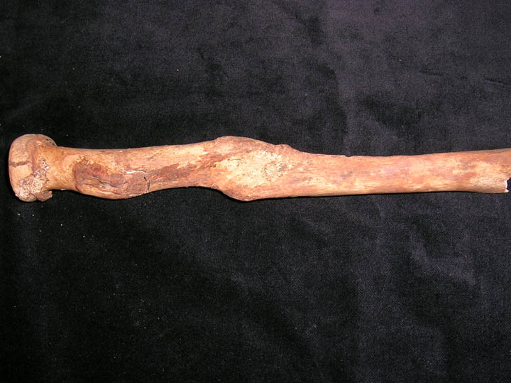
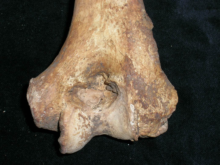
Osteoarthritis of carpals and metacarpal
|
| OCU00
|
35
|
5
|
OCU00_35_5.jpg
|
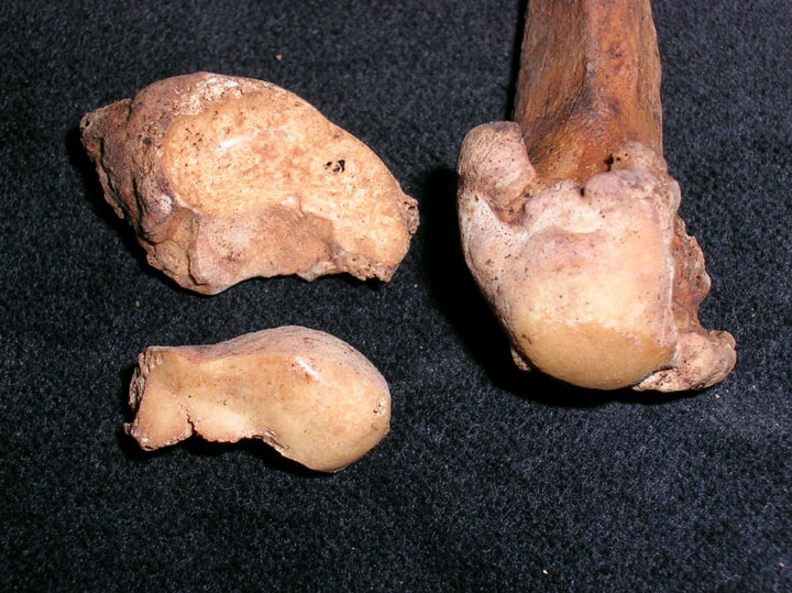
Osteoarthritis of carpals
|
| OCU00
|
35
|
6
|
OCU00_35_6.jpg
|
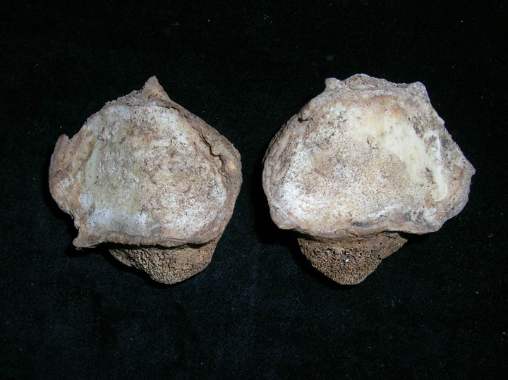
Bilateral eburnation of the distal radii
|
| OCU00
|
35
|
7
|
OCU00_35_7.jpg
|
Destructive lesions on distal aspect of MT1(Gout) (Dorsal view)
|
| OCU00
|
35
|
8
|
OCU00_35_8.jpg
|
Destructive lesions on distal aspect of MT1(Gout) (Lateral view)
|
| OCU00
|
39
|
1
|
OCU00_39_1.jpg
|
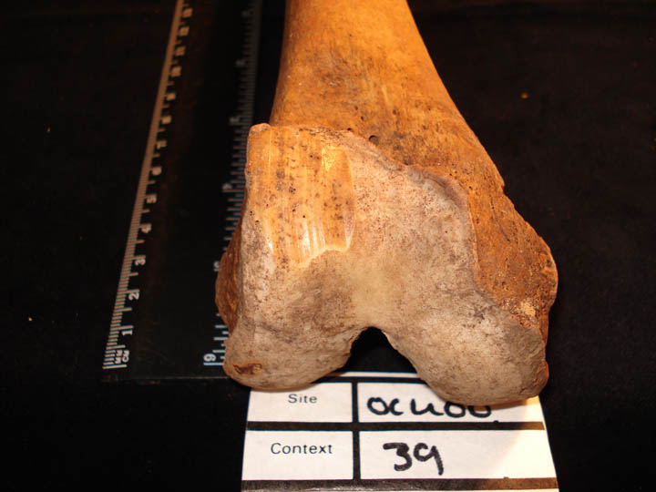
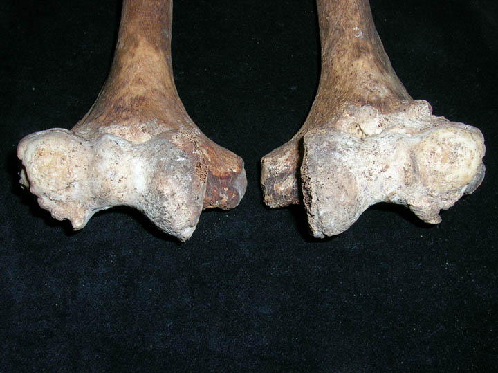
Bilateral osteoarthritis of the femora with eburnation of the anterior joint surface and grooving on the right anterior joint surface
|
| OCU00
|
39
|
2
|
OCU00_39_2.jpg
|
Osteoarthritis of the right femur showing eburnation and grooving of the
|
| OCU00
|
39
|
3
|
OCU00_39_3.jpg
|
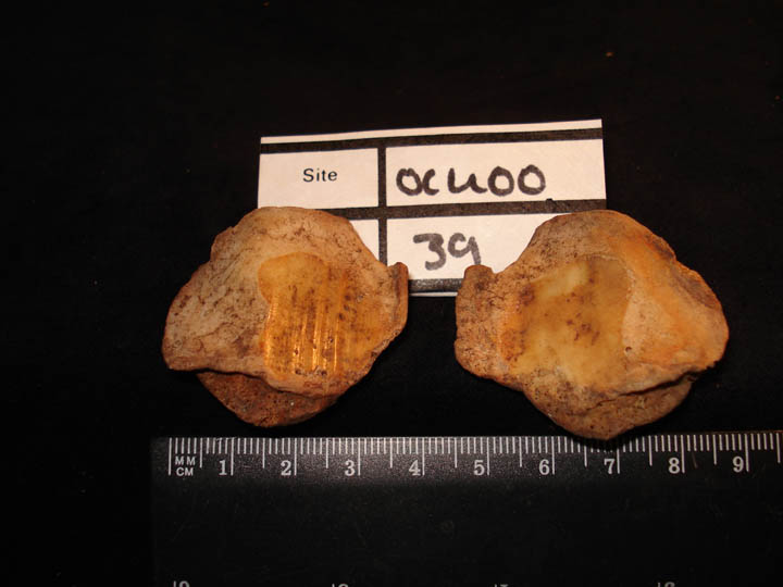
Osteoarthtritis of the patellae (posterior view) showing eburnation and grooving of the right side
|
| OCU00
|
39
|
4
|
OCU00_39_4.jpg
|
Patellae (anterior view) showing bilateral vastus notch
|
| OCU00
|
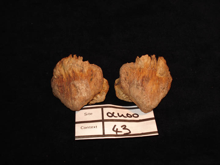
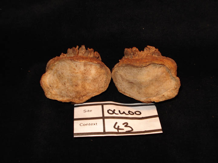
43
|
1
|
OCU00_43_1.jpg
|
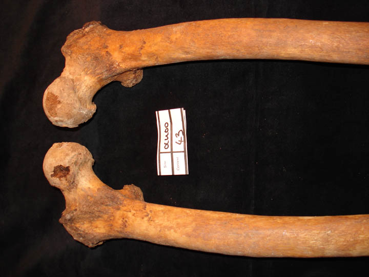
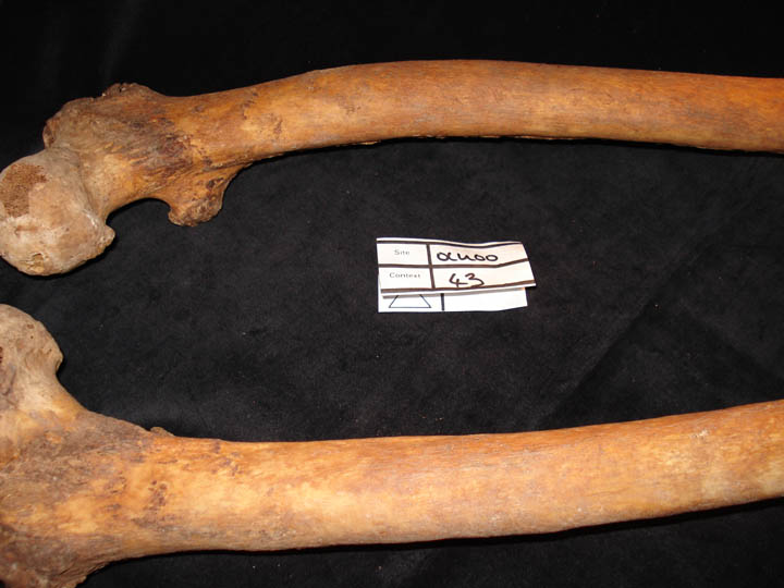
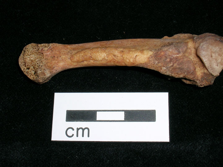
Residual/healed rickets of the femora
|
| OCU00
|
43
|
2
|
OCU00_43_2.jpg
|
Residual/healed rickets of the femora
|
| OCU00
|
43
|
3
|
OCU00_43_3.jpg
|
Patellae (anterior view) showing pronounced enthesophytic development of Rectus femoris
|
| OCU00
|
43
|
4
|
OCU00_43_4.jpg
|
Patellae (posterior view) showing marginal osteophytic lipping and enthesophytic development
|
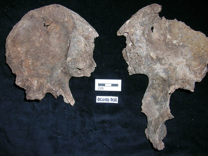
| OCU00
|
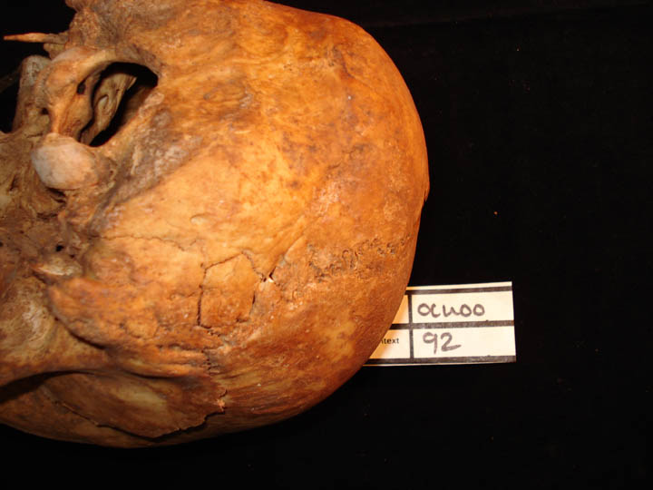
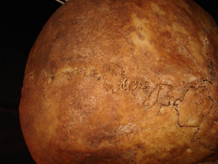
92
|
1
|
OCU00_92_1.jpg
|
Asterionic bone right side
|
| OCU00
|
92
|
2
|
OCU00_92_2.jpg
|
Asterionic bone right side
|
| OCU00
|
92
|
3
|
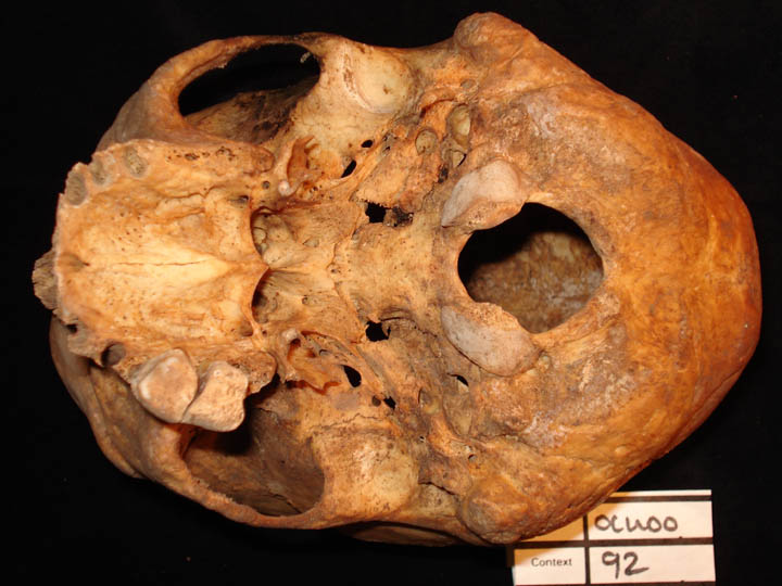
OCU00_92_3.jpg
|
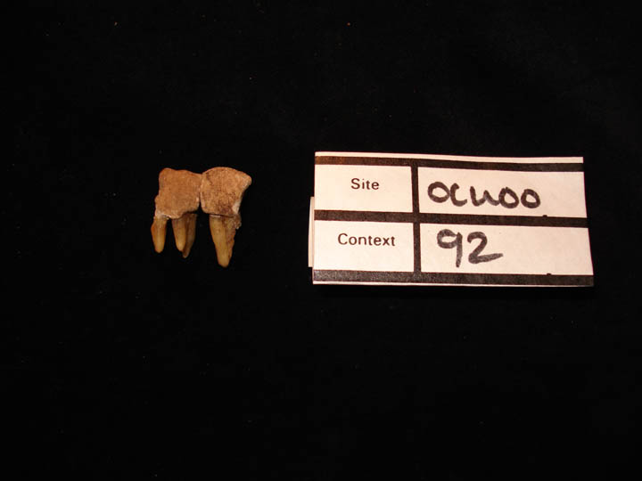
Severe calculus Grade 3 of the right 1st & 2nd maxillary molars (buccal view)
|
| OCU00
|
92
|
4
|
OCU00_92_4.jpg
|
Severe calculus Grade 3 of the right 1st & 2nd maxillary molars (buccal view)
|
| OCU00
|
92
|
5
|
OCU00_92_5.jpg
|
Severe calculus Grade 3 of the right 1st & 2nd maxillary molars (buccal view)
|
| OCU00
|
92
|
6
|
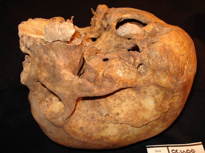
OCU00_92_6.jpg
|
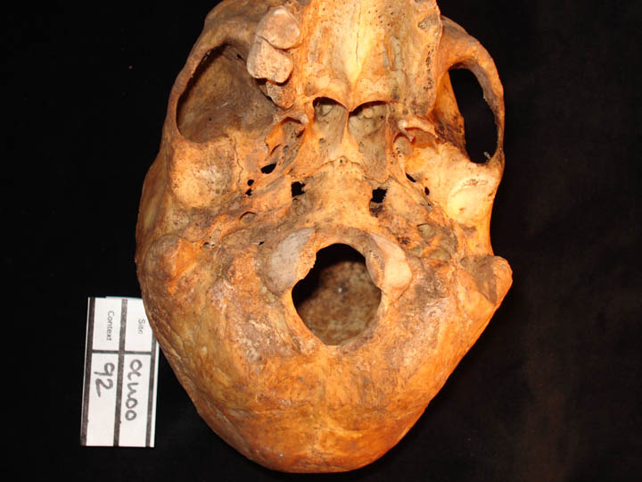
Base of skull showing the tempormandibular joints and the change to the right side shallow and flattened (posterior view)
|
| OCU00
|
92
|
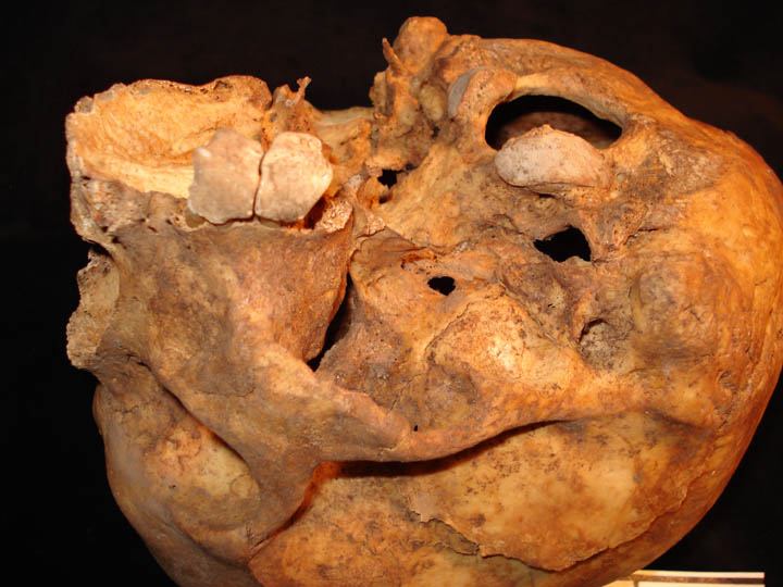
7
|
OCU00_92_7.jpg
|
Base of skull showing the tempormandibular joints and change to the right side shallow and flattened (posterior view)
|
| OCU00
|
92
|
8
|
OCU00_92_8.jpg
|
Maxillary molars 1st and 2nd showing severe calculus (Grade 3) covering all the enamel
|
| OCU00
|
115
|
1
|
OCU00_115_1.jpg
|
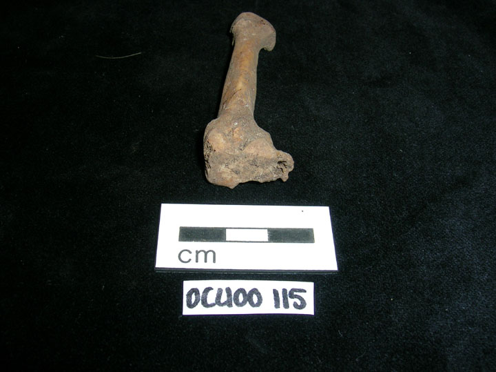
Metatarsal pitting of MT3
|
| OCU00
|
115
|
2
|
OCU00_115_2.jpg
|
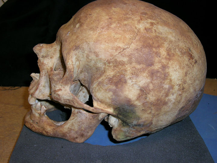
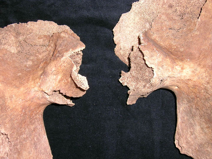
Anterior superior view of skull with circular defect (Postmortem?)
|
| OCU00
|
115
|
3
|
OCU00_115_3.jpg
|
Left lateral view of skull with nasal fracture and antemorem tooth loss
|
| OCU00
|
115
|
4
|
OCU00_115_4.jpg
|
Nasal Fracture
|
| OCU00
|
115
|
5
|
OCU00_115_5.jpg
|
Tarsal coalision of the calcaneum and navicular
|
| OCU00
|
147
|
1
|
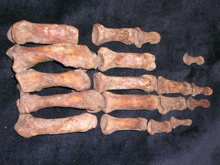
OCU00_147_1.jpg
|
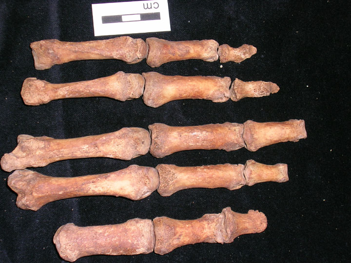
Left hand
|
| OCU00
|
147
|
2
|
OCU00_147_2.jpg
|
Osteoarthritis of the IPJ
|
| OCU00
|
147
|
3
|
OCU00_147_3.jpg
|
Osteoarthritis of the IPJ
|
| OCU00
|
147
|
4
|
OCU00_147_4.jpg
|
Periosteal new bone growth on MT4
|
| OCU00
|
147
|
5
|
OCU00_147_5.jpg
|
Ankylosis of phlangeal joint
|
| OCU00
|
147
|
6
|
OCU00_147_6.jpg
|
Fusion of tibia and fibula follwing trauma
|
| OCU00
|
147
|
7
|
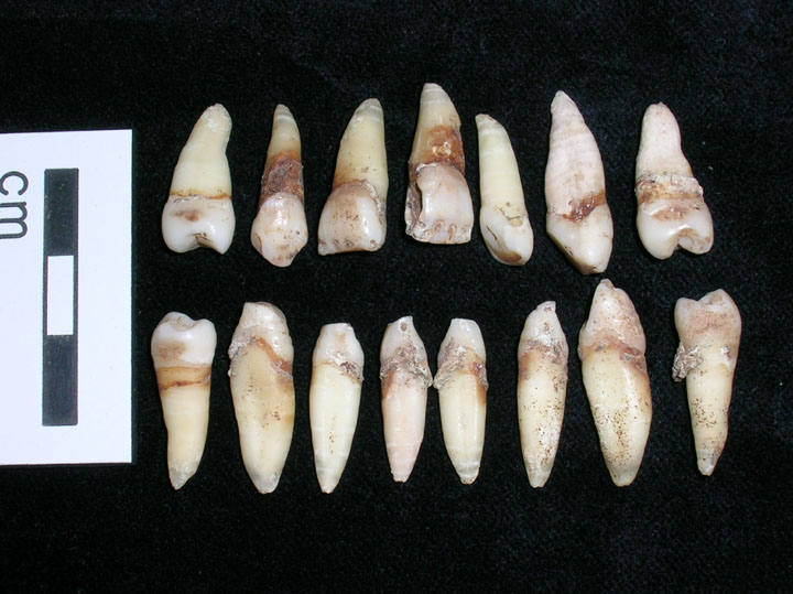
OCU00_147_7.jpg
|
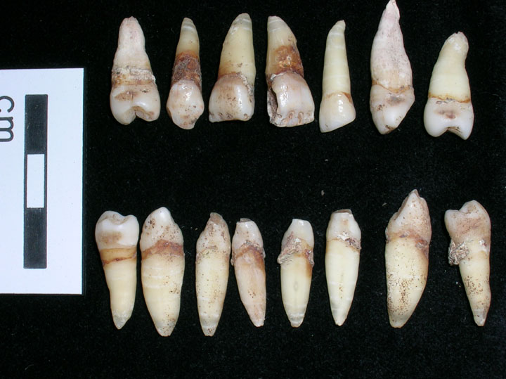
Dentition displaying root banding
|
| OCU00
|
147
|
8
|
OCU00_147_8.jpg
|
Mandibular dentition displaying root banding
|
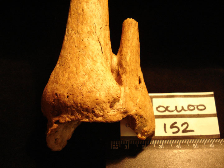
| OCU00
|
152
|
1
|
OCU00_152_1.jpg
|
Healed fracture of the distal end of the left tibia and fibula with ankylosis (anterior view)
|
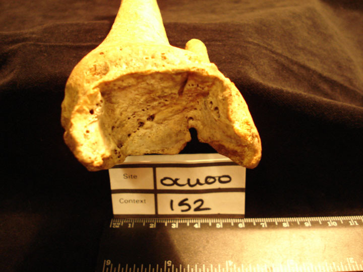
| OCU00
|
152
|
2
|
OCU00_152_2.jpg
|
Healed fracture of the distal end of the left tibia and fibula with ankylosis showing the talocrual articular surface with fracture line and secondary joint changes (posterior view)
|
| OCU00
|
154
|
1
|
OCU00_154_1.jpg
|
Concha Bulbosa of the left concha
|
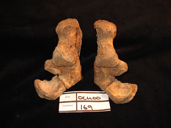
| OCU00
|
169
|
1
|
OCU00_169_1.jpg
|
Left and right calcanea showing tarsal coalition (calcaneonavicular)
|
| OCU00
|
188
|
1
|
OCU00_188_1.jpg
|
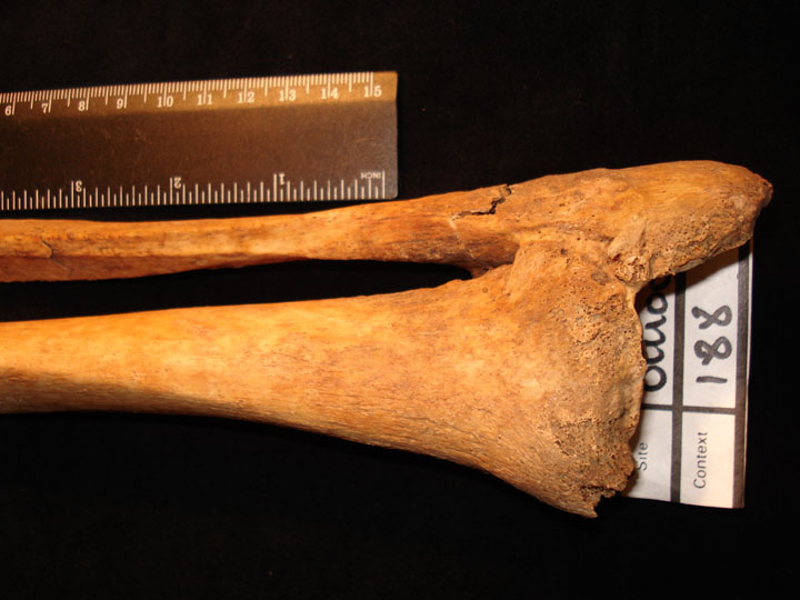
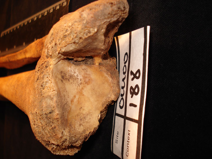
Ankylosis of the left tibiofibula joint (anterior view)
|
| OCU00
|
188
|
2
|
OCU00_188_2.jpg
|
Ankylosis of the left tibiofibula joint (anterior view)
|
| OCU00
|
198
|
1
|
OCU00_198_1.jpg
|
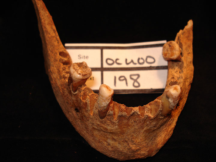
Hypoplastic defects of mandibular molars
|
| OCU00
|
198
|
2
|
OCU00_198_2.jpg
|
Hypoplastic defects of mandibular molars
|
| OCU00
|
198
|
3
|
OCU00_198_3.jpg
|
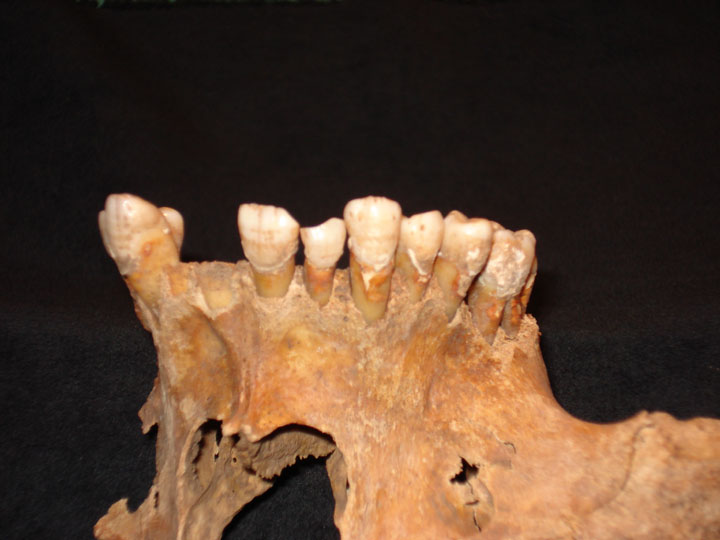
Maxillary teeth right central and lateral incisors exhibiting pipe facets
|
| OCU00
|
198
|
4
|
OCU00_198_4.jpg
|
Mandibular teeth right canine and lateal incisor exhibiting pipe facets
|
| OCU00
|
206
|
1
|
OCU00_206_1.jpg
|
Anterior mandibular dentition with hypoplastic defects on the enamel
|
| OCU00
|
206
|
2
|
OCU00_206_2.jpg
|
Anterior maxillary dentition with hypoplastic defects
|
| OCU00
|
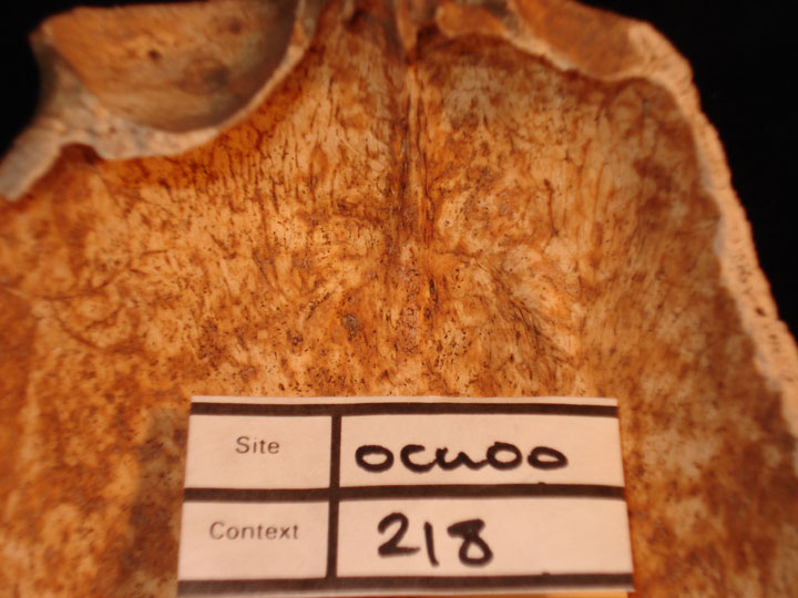
218
|
1
|
OCU00_218_1.jpg
|
Skull Frontal bone endocranial surface showing nodular changes associated with hyperostosis frontalis interna
|
| OCU00
|
218
|
2
|
OCU00_218_2.jpg
|
Skull Frontal bone endocranial surface showing nodular changes associated with hyperostosis frontalis interna
|
| OCU00
|
230
|
1
|
OCU00_230_1.jpg
|
Anterior maxillary dentition with hypoplastic defects (sub adult)
|
| OCU00
|
230
|
2
|
OCU00_230_2.jpg
|
Maxillary dentition with dental enamel hypoplastic defects (sub adult)
|
| OCU00
|
230
|
3
|
OCU00_230_3.jpg
|
Mandibular dentition with dental enamel hypoplastic defect
|
| OCU00
|
232
|
1
|
OCU00_232_1.jpg
|
Right hand with fracture of MC2
|
| OCU00
|
258
|
1
|
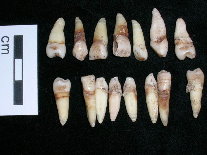
OCU00_258_1.jpg
|
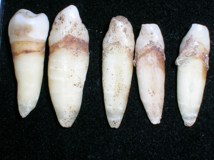
Dentition displaying root banding
|
| OCU00
|
258
|
2
|
OCU00_258_2.jpg
|
Dentition displaying root banding
|
| OCU00
|
339
|
1
|
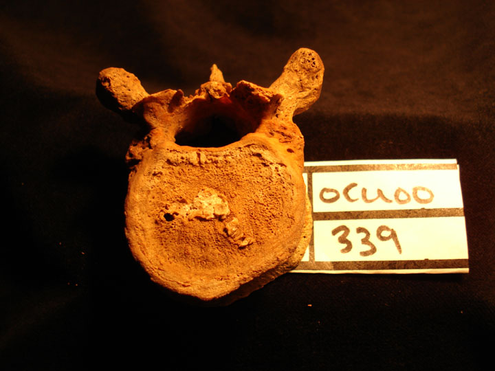
OCU00_339_1.jpg
|
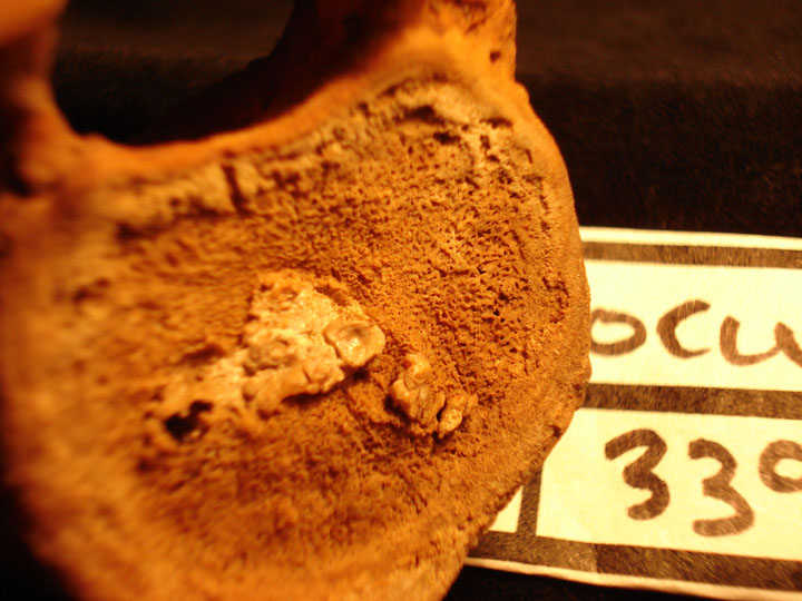
Thoracic vertebra TH11 showing possible destructive lesion of the centrum (?TB) with calcified cyst/pus (superior view)
|
| OCU00
|
339
|
2
|
OCU00_339_2.jpg
|
Thoracic vertebra TH11 showing possible destructive lesion of the centrum (?TB) with calcified cyst/pus (superior view)
|
| OCU00
|
339
|
3
|
OCU00_339_3.jpg
|
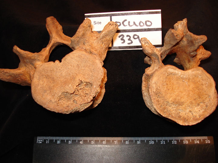
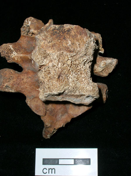
Thoracic vertebra TH12 showing possible destructive lesion of the centrum and annulus fibrosus (?TB) (inferior view) and Lumbar vertebra L5 (superior view) showing destruction of corresponding vertebral area
|
| OCU00
|
339
|
4
|
OCU00_339_4.jpg
|
Thoracic vertebra TH12 showing possible destructive lesion of the centrum and annulus fibrosus (?TB) (inferior view) and Lumbar vertebra L5 (superior view) showing destruction of corresponding vertebral area
|
| OCU00
|
339
|
5
|
OCU00_339_5.jpg
|
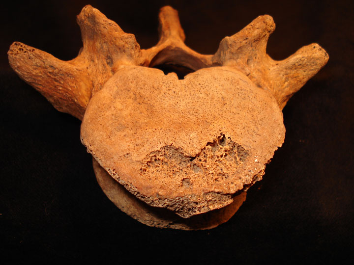
Lumbar L5 (superior view) showing area of destruction possibly lytic ?TB
|
| OCU00
|
339
|
6
|
OCU00_339_6.jpg
|
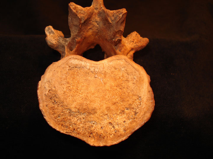
Thoracic TH12 (inferior view) showing area of destrction [possibly lytic ?TB
|
| OCU00
|
343
|
1
|
OCU00_343_1.jpg
|
Ankylosis of left sacroilliac joint with distortion (anterior view)
|
| OCU00
|
343
|
2
|
OCU00_343_2.jpg
|
Ankylosis of left sacroilliac joint with distortion (posterior view)
|
| OCU00
|
343
|
3
|
OCU00_343_3.jpg
|
Ankylosis of left sacroilliac joint with distortion (posterior view)
|
| OCU00
|
343
|
4
|
OCU00_343_4.jpg
|
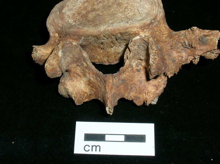
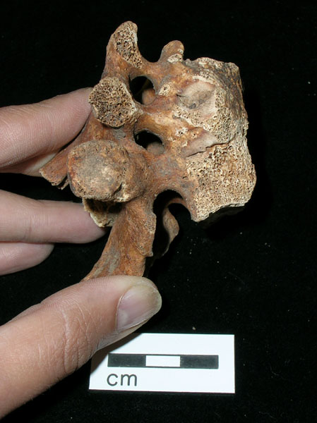
6th lumbar vertebra diplaying wedging and changes to inferior facets (inferior view)
|
| OCU00
|
343
|
5
|
OCU00_343_5.jpg
|
6th lumbar vertebra diplaying wedging and changes to inferior facets (posterior view)
|
| OCU00
|
343
|
6
|
OCU00_343_6.jpg
|
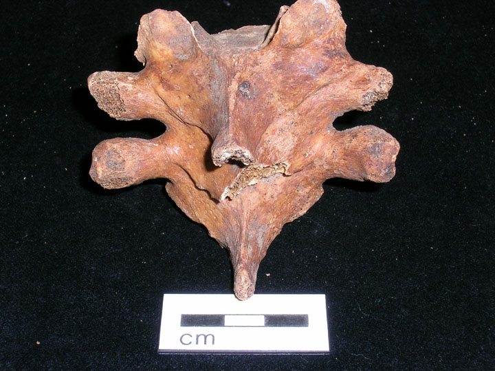
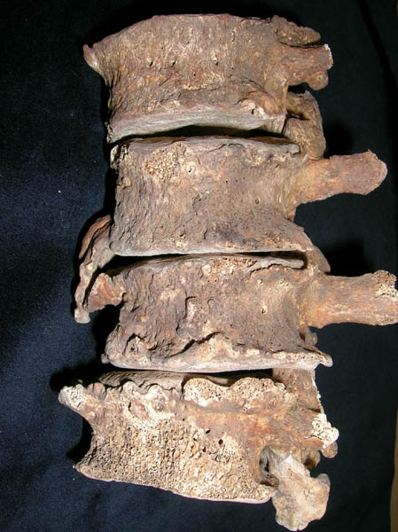
Fusion of cervical and thoracic vertebrae (anterior view)
|
| OCU00
|
343
|
7
|
OCU00_343_7.jpg
|
Fusion of cervical and thoracic vertebrae (Lateral view)
|
| OCU00
|
343
|
8
|
OCU00_343_8.jpg
|
Fusion and compression of cervical and thoracic veretebrae (posterior view)
|
| OCU00
|
349
|
1
|
OCU00_349_1.jpg
|
Anterior mandibular dentition with hypoplastic defects on the enamel
|
| OCU00
|
349
|
2
|
OCU00_349_2.jpg
|
Anterior dentition with hypoplastic defects of the enamel
|
| OCU00
|
349
|
3
|
OCU00_349_3.jpg
|
Anterior view of skull with dental enamel hypoplasia
|
| OCU00
|
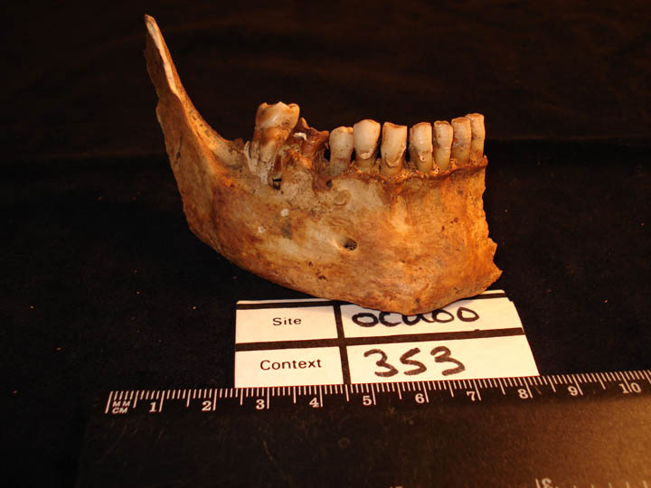
353
|
1
|
OCU00_353_1.jpg
|
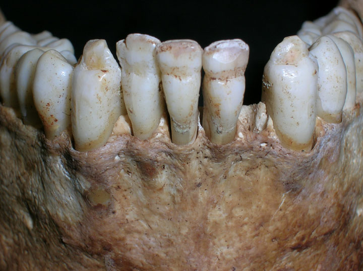
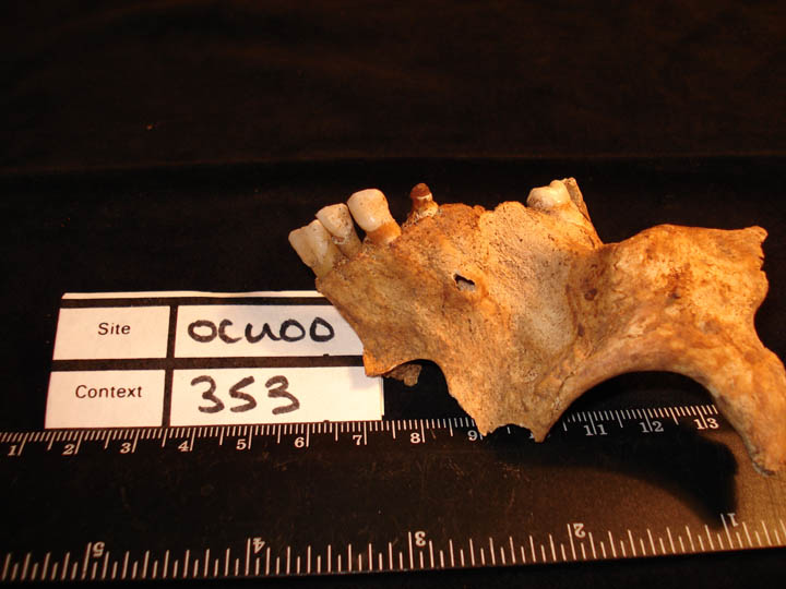
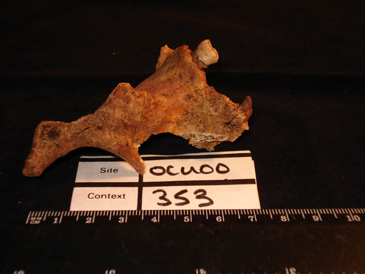
Carious destruction of right maxillary PM3
|
| OCU00
|
353
|
2
|
OCU00_353_2.jpg
|
Carious destruction of left maxillary PM3
|
| OCU00
|
353
|
3
|
OCU00_353_3.jpg
|
Carious destrution of right mandibular 1st molar
|
| OCU00
|
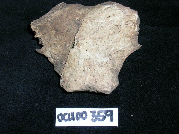
359
|
1
|
OCU00_359_1.jpg
|
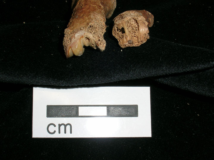
Diagonal dissection of manubrium
|
| OCU00
|
363
|
1
|
OCU00_363_1.jpg
|
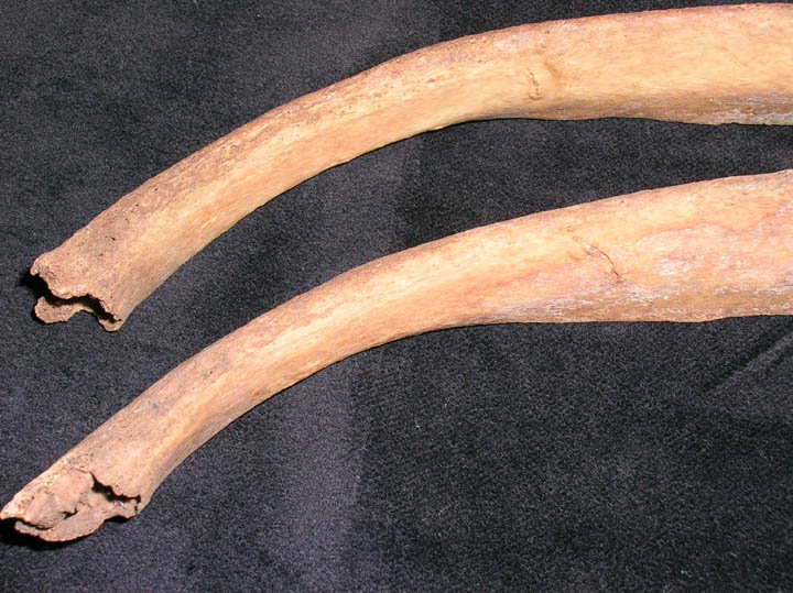
Bowing deformity 'non plastic bending' of the right tibia and fibula (with left as comparison)
|
| OCU00
|
383
|
1
|
OCU00_383_1.jpg
|
Spina bifida occulta in sub adult
|
| OCU00
|
392
|
1
|
OCU00_392_1.jpg
|
Frontal bone with periosteal changes
|
| OCU00
|
392
|
2
|
OCU00_392_2.jpg
|
Frontal bone with periosteal changes (close-up)
|
| OCU00
|
392
|
3
|
OCU00_392_3.jpg
|
Left clavicle with periosteal changes
|
| OCU00
|
392
|
4
|
OCU00_392_4.jpg
|
Acromioclavicular joint of left clavicel with erosive lesions
|
| OCU00
|
392
|
5
|
OCU00_392_5.jpg
|
Periosteal new bone of distal left femur
|
| OCU00
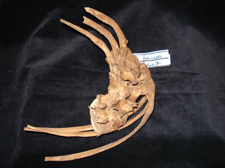
|
407
|
1
|
OCU00_407_1.jpg
|
Kyphoscoloiosis with involvement of ribs (posterior view)
|
| OCU00
|
407
|
2
|
OCU00_407_2.jpg
|
Kyphoscoloiosis with involvement of ribs (anterior view)
|
| OCU00
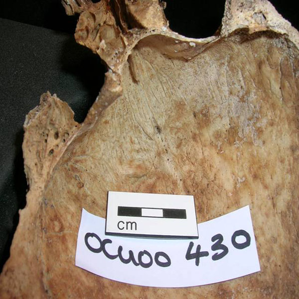
|
430
|
1
|
OCU00_430_1.jpg
|
Endocranial surface showing changes associated to hyperostosis frontalis interna (HFI) Stage 2
|
| OCU00
|
436
|
1
|
OCU00_436_1.jpg
|
Myositis ossificans on proximal left tibia
|
| OCU00
|
436
|
2
|
OCU00_436_2.jpg
|
Osteoarthritis of lateral border of femorapatellar joint of left femur
|
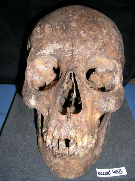
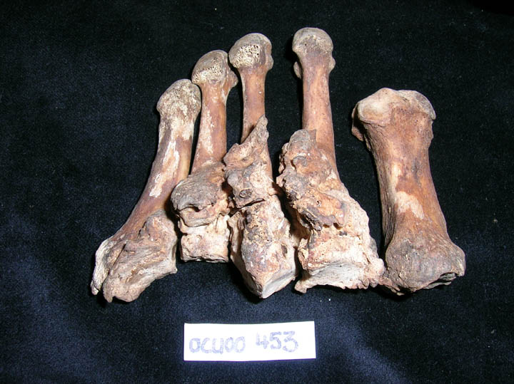
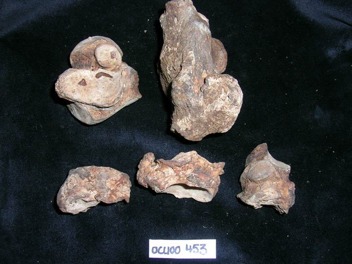
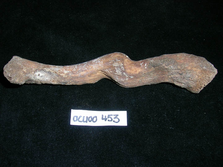
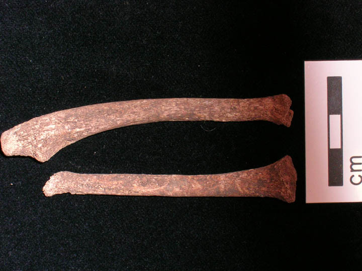
| OCU00
|
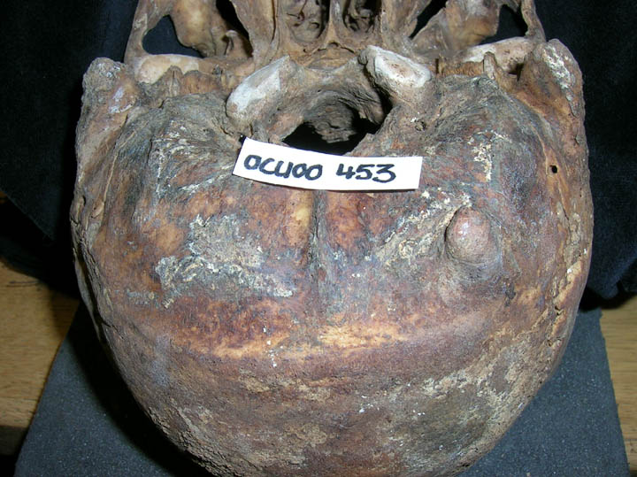
453
|
1
|
OCU00_453_1.jpg
|
Maxillary dentition with caries and uneven wear pattern
|
| OCU00
|
453
|
2
|
OCU00_453_2.jpg
|
Bony exostosis on left occipital bone
|
| OCU00
|
453
|
3
|
OCU00_453_3.jpg
|
Anterior view of skull with marked gap in anterior dentition
|
| OCU00
|
453
|
4
|
OCU00_453_4.jpg
|
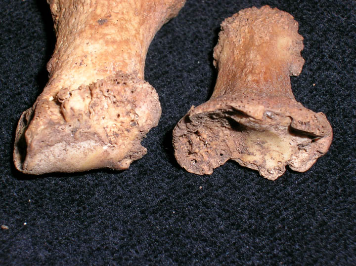
Gross exostosis on plantar aspect of right foot
|
| OCU00
|
453
|
5
|
OCU00_453_5.jpg
|
Tarsal bony outgrowth
|
| OCU00
|
453
|
6
|
OCU00_453_6.jpg
|
Fracture of right clavicle
|
| OCU00
|
456
|
1
|
OCU00_456_1.jpg
|
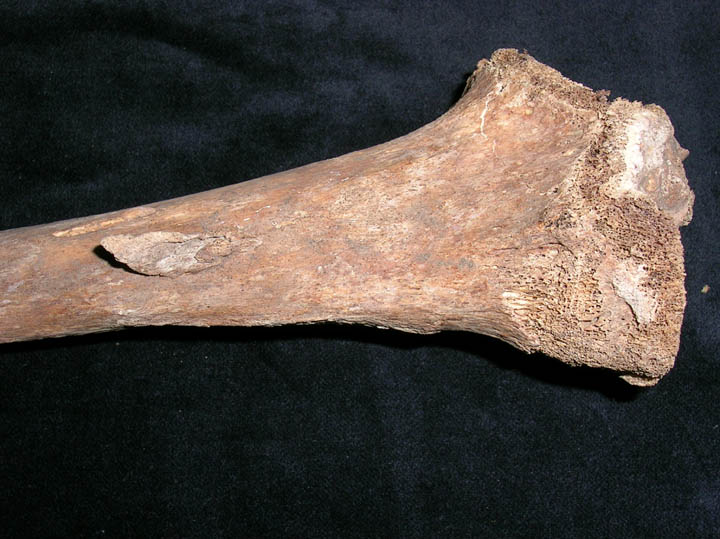
Active rickets on right distal radius and ulna
|
| OCU00
|
456
|
2
|
OCU00_456_2.jpg
|
Active rickets on right distal ulna
|
| OCU00
|
456
|
3
|
OCU00_456_3.jpg
|
Active rickets on right distal radius
|
| OCU00
|
456
|
4
|
OCU00_456_4.jpg
|
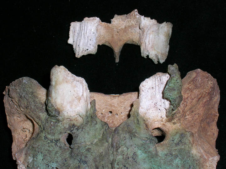
Active rickets shown as flaring of the sternal end of ribs
|
| OCU00
|
456
|
5
|
OCU00_456_5.jpg
|
Active rickets shown as flaring of the sternal end of rib
|
| OCU00
|
456
|
6
|
OCU00_456_6.jpg
|
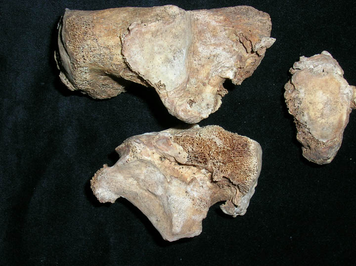
Active rickets of femora with flattening of proximal epihyses and flaring of distal epiphyses
|
| OCU00
|
456
|
7
|
OCU00_456_7.jpg
|
Active rickets of tibia with angulation of the distal epiphysis
|
| OCU00
|
456
|
8
|
OCU00_456_8.jpg
|
Active rickets of tibia with angulation of the distal epiphysis
|
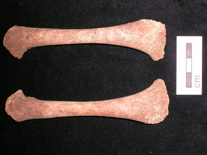
| OCU00
|
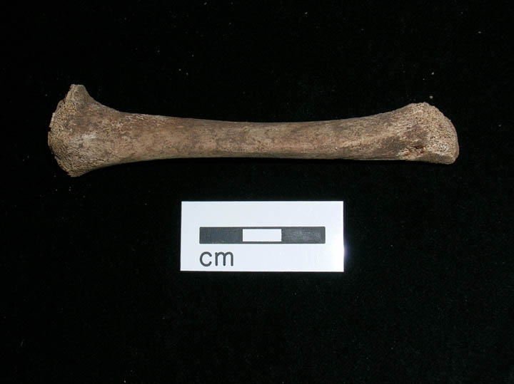
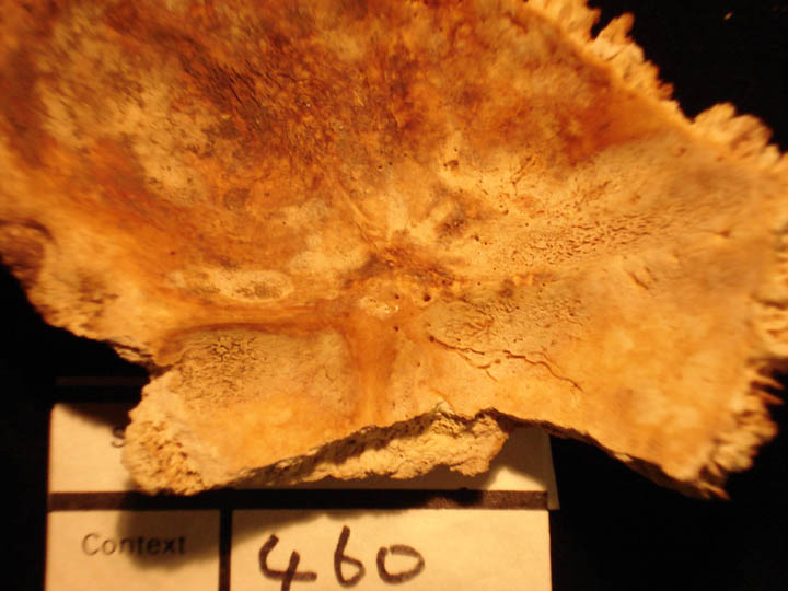
460
|
1
|
OCU00_460_1.jpg
|
Occipital bone endocranial surface increase in porosity with labyrinthine bone formation
|
| OCU00
|
474
|
1
|
OCU00_474_1.jpg
|
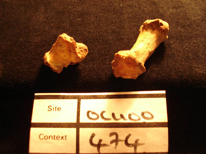
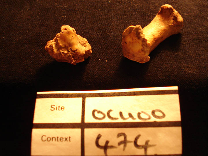
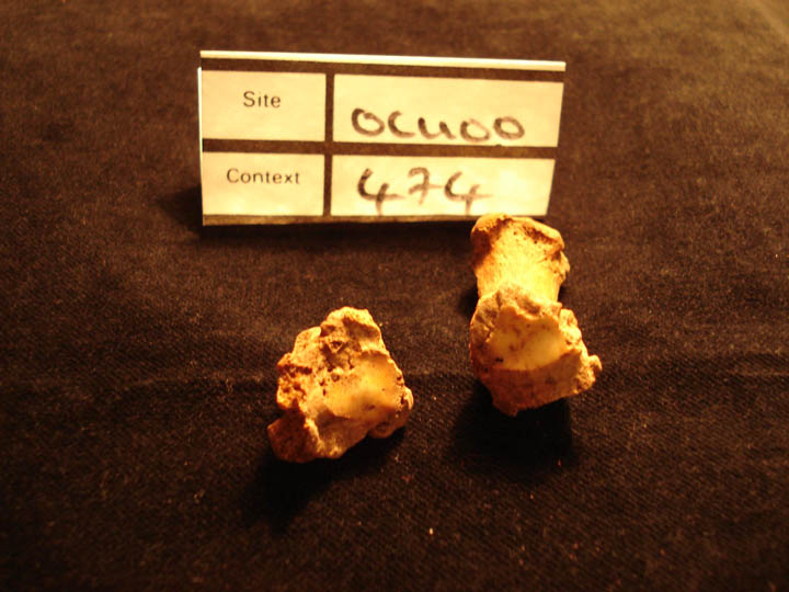
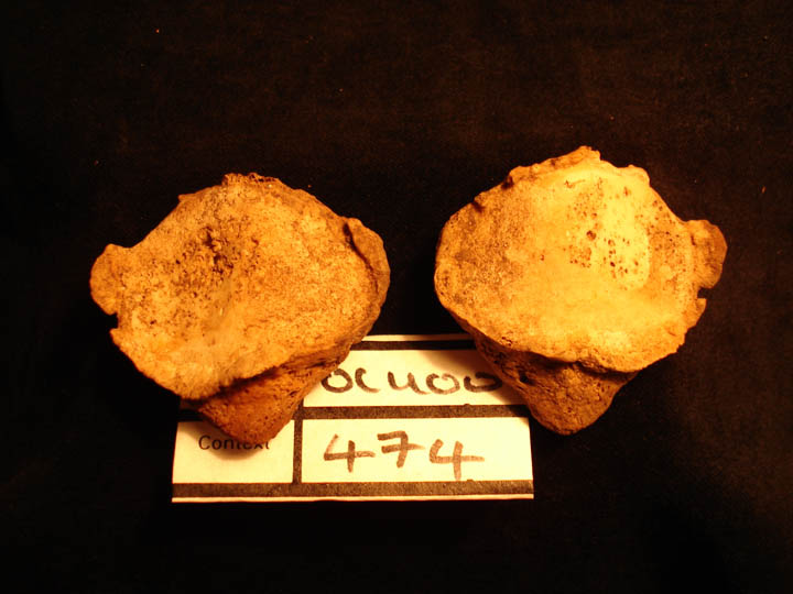
Right 1st metacarpal and trapezium showing osteoarthritis of the joint surfaces and eburnation
|
| OCU00
|
474
|
2
|
OCU00_474_2.jpg
|
Left 1st metacarpal and trapezium showing osteoarthritis of the joint surfaces and eburnation
|
| OCU00
|
474
|
3
|
OCU00_474_3.jpg
|
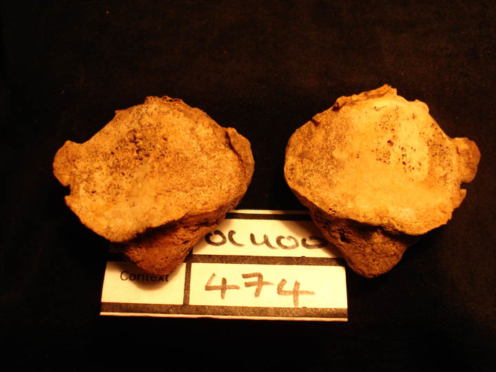
Left 1st metacarpal and trapezium showing osteoarthritis and gross changes of the joint surfaces and eburnation
|
| OCU00
|
474
|
4
|
OCU00_474_4.jpg
|
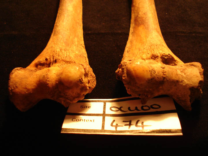
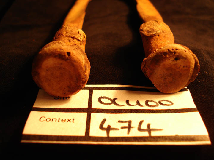
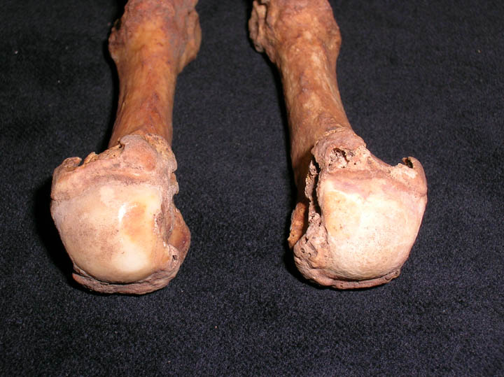
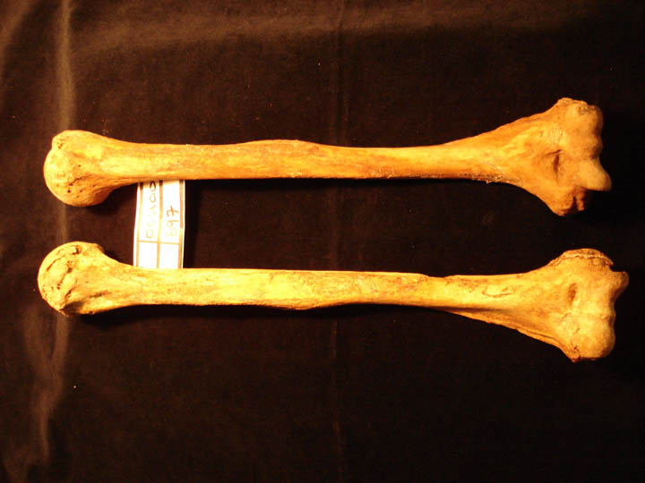
Humerii showing bilateral osteoarthritis and eburnation of the humeroradial joint surfaces (anterior view)
|
| OCU00
|
474
|
5
|
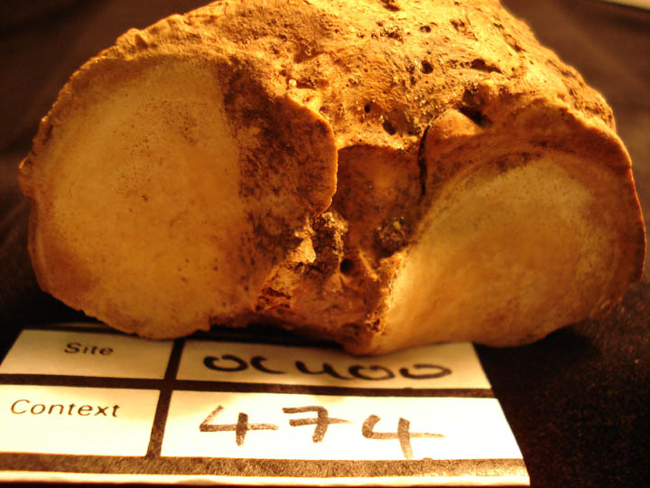
OCU00_474_5.jpg
|
Radii showing bilateral osteoarthritis and eburnation of the radial heads (superior view)
|
| OCU00
|
474
|
6
|
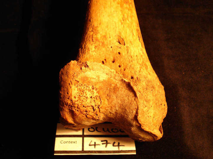
OCU00_474_6.jpg
|
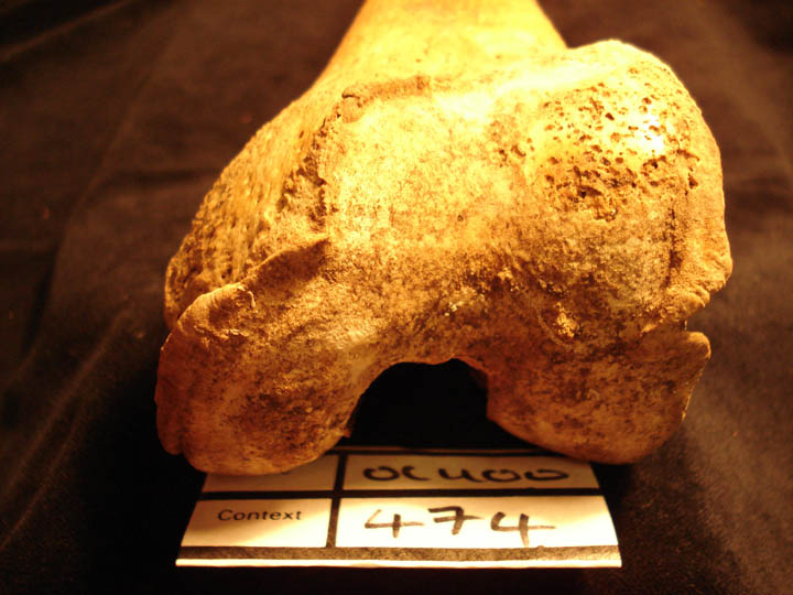
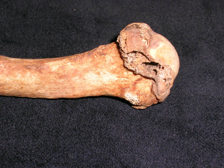
Right femur showing osteoarthrits of the anterior joint surface with destruction and eburnation (anteior view)
|
| OCU00
|
474
|
7
|
OCU00_474_7.jpg
|
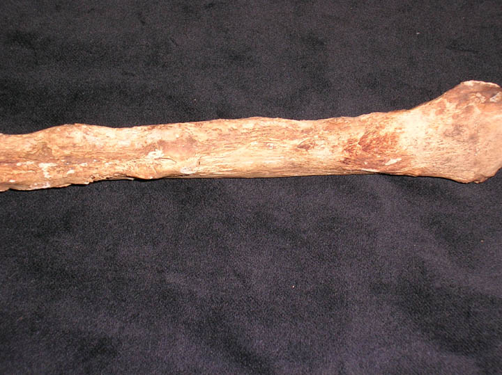
Right tibia showing osteophytic marginal lipping of the medial and lateral joint surfaces (superior view)
|
| OCU00
|
474
|
8
|
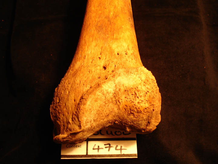
OCU00_474_8.jpg
|
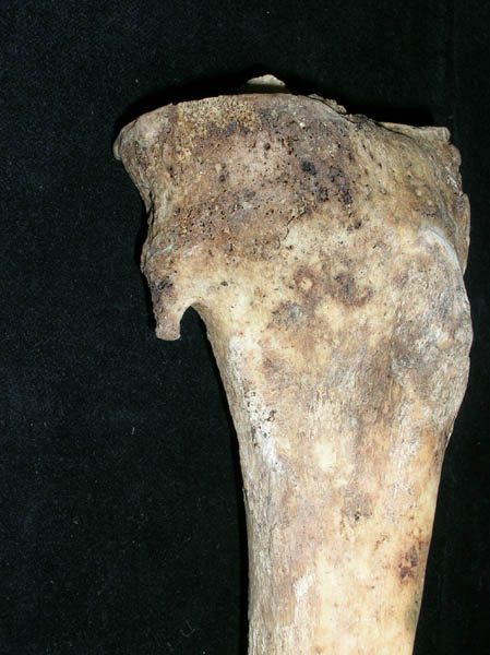
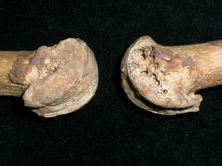
Left femur showing osteoarthritis of the anterior joint surface with destruction and eburnation (anterior view)
|
| OCU00
|
474
|
9
|
OCU00_474_9.jpg
|
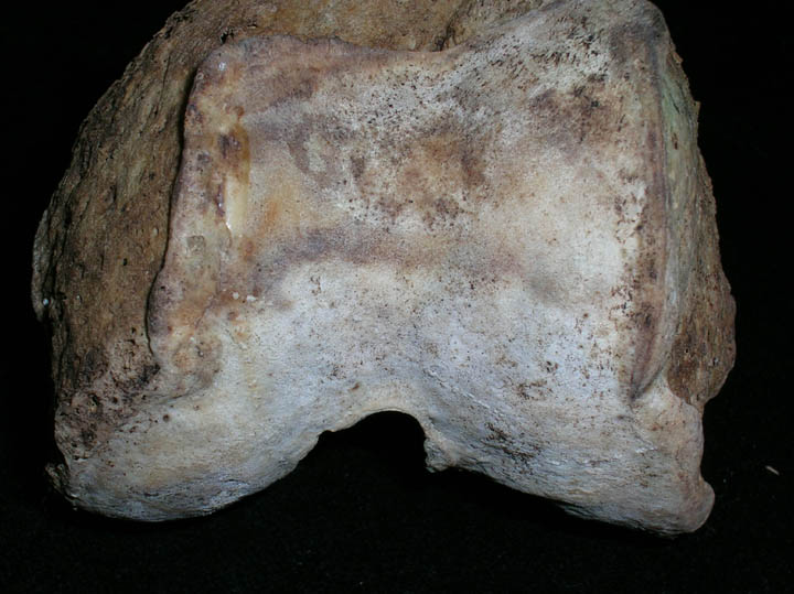
Left femur showing (close up) osteoarthritis of the anterior joint surface with destruction and eburnation (anterior view)
|
| OCU00
|
474
|
10
|
OCU00_474_10.jpg
|
Patellae showing osteoarthritis with marginal lipping and eburnation (posteior view)
|
| OCU00
|
474
|
11
|
OCU00_474_11.jpg
|
Patellae showing osteoarthritis with marginal lipping and eburnation (posteior view)
|
| OCU00
|
474
|
12
|
OCU00_474_12.jpg
|
Sternum with xyphoid process
|
| OCU00
|
483
|
1
|
OCU00_483_1.jpg
|
Edentulous mandible
|
| OCU00
|
483
|
2
|
OCU00_483_2.jpg
|
Circular smooth edged defects of the sphenoid (left side/inferior view)
|
| OCU00
|
494
|
1
|
OCU00_494_1.jpg
|
Sacrum showing defect of S5 to S2 (posterior view)
|
| OCU00
|
494
|
2
|
OCU00_494_2.jpg
|
Severe osteophytic lipping of lumbar vertebrae (anterior view)
|
| OCU00
|
494
|
3
|
OCU00_494_3.jpg
|
Severe osteophytic lipping of lumbar vertebrae (anterior view)
|
| OCU00
|
496
|
1
|
OCU00_496_1.jpg
|
Complete thyroid
|
| OCU00
|
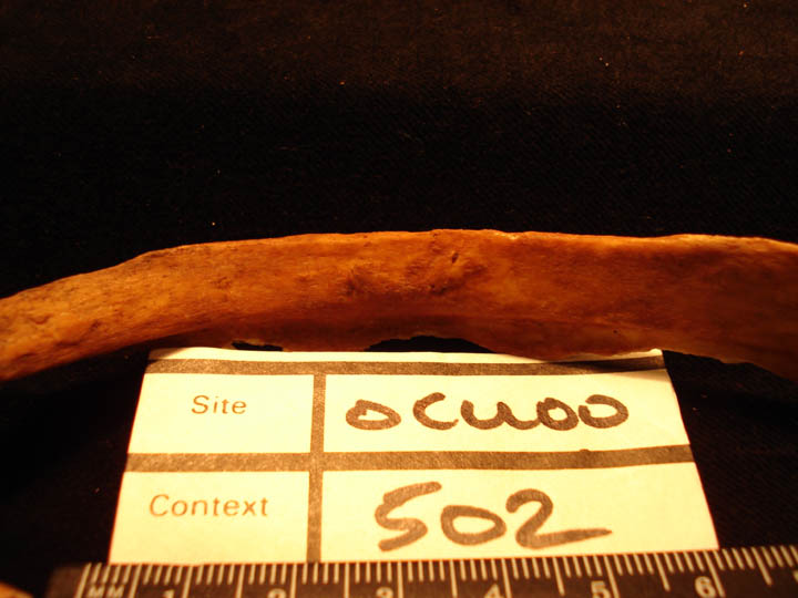
502
|
1
|
OCU00_502_1.jpg
|
Rib lesion on the visceral surface, healed and remodelled
|
| OCU00
|
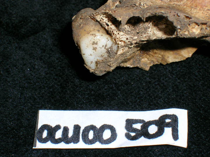
509
|
1
|
OCU00_509_1.jpg
|
Impacted dentition
|
| OCU00
|
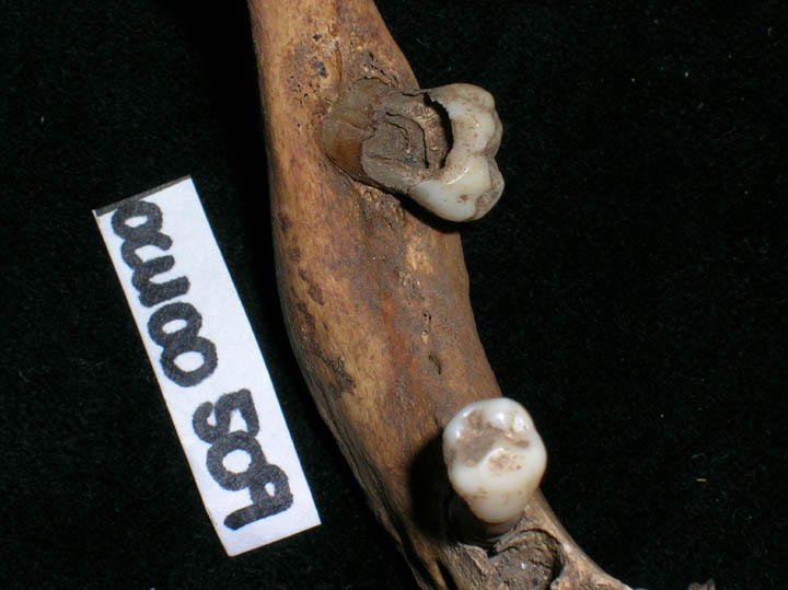
509
|
2
|
OCU00_509_2.jpg
|
Caries and antemortem tooth loss in mandible
|
| OCU00
|
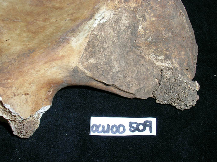
509
|
3
|
OCU00_509_3.jpg
|
Ankylosis along inferior margin of the right illium
|
| OCU00
|
525
|
1
|
OCU00_525_1.jpg
|
DISH (right lateral view)
|
| OCU00
|
525
|
2
|
OCU00_525_2.jpg
|
DISH (Left lateral view)
|
| OCU00
|
525
|
3
|
OCU00_525_3.jpg
|
Bilateral osteoarthritis of ulnae
|
| OCU00
|
525
|
4
|
OCU00_525_4.jpg
|
Ankylosis of right sacroilliac joint
|
| OCU00
|
525
|
5
|
OCU00_525_5.jpg
|
Bilateral osteoarthritis of patellae
|
| OCU00
|
525
|
6
|
OCU00_525_6.jpg
|
Bilateral osteoarthritis of distal humerii
|
| OCU00
|
525
|
7
|
OCU00_525_7.jpg
|
Edentulous anterior view of skull
|
| OCU00
|
525
|
8
|
OCU00_525_8.jpg
|
Edentulous left lateral view of skull
|
| OCU00
|
527
|
1
|
OCU00_527_1.jpg
|
Congenital ankylosis of right ribs (bifid rib)
|
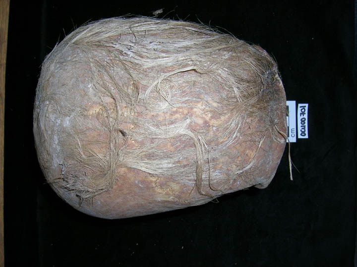
| OCU00
|
534
|
1
|
OCU00_534_1.jpg
|
Superior view of skull with hair
|
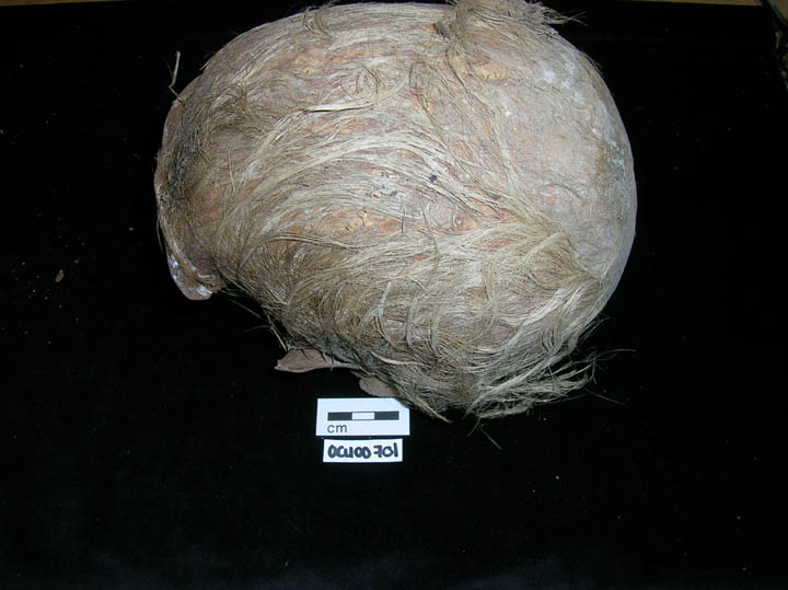
| OCU00
|
552
|
1
|
OCU00_552_1.jpg
|
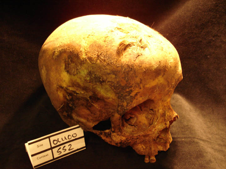
Skull of older female showing preserved hair on the frontal bone
|
| OCU00
|
552
|
2
|
OCU00_552_2.jpg
|
Skull of older female showing preserved hair on the frontal bone
|
| OCU00
|
562
|
1
|
OCU00_562_1.jpg
|
Osteoarthritis of the left scaphoid and lunate showing eburnated articular surfaces
|
| OCU00
|
562
|
2
|
OCU00_562_2.jpg
|
Osteoarthritis of the left scaphoid and lunate showing eburnated articular surfaces
|
| OCU00
|
562
|
3
|
OCU00_562_3.jpg
|
Osteoarthritis of the left capitate showing eburnation to the capitate head
|
| OCU00
|
579
|
1
|
OCU00_579_1.jpg
|
Depression (blunt force trauma?) on left frontal bone
|
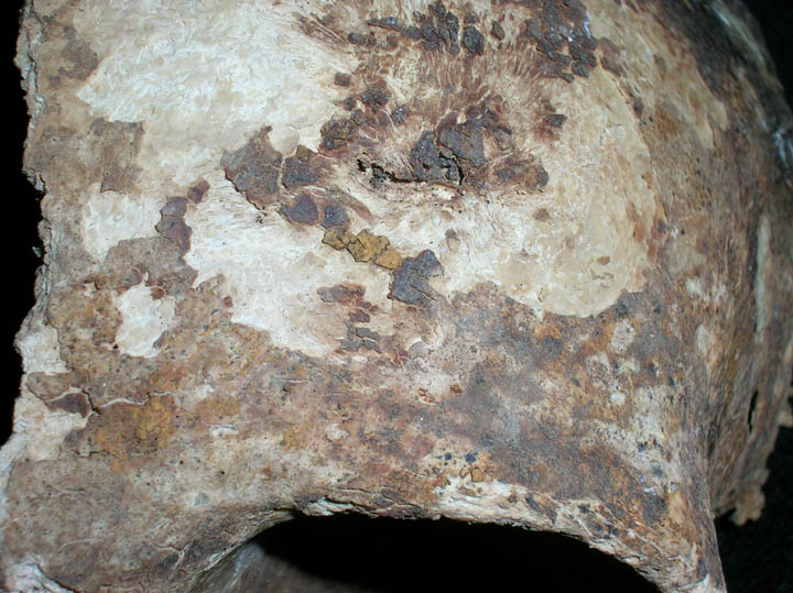
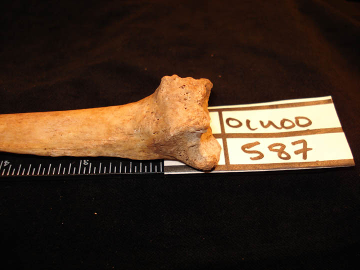
| OCU00
|
587
|
1
|
OCU00_587_1.jpg
|
Healed Colles fracture of the distal end of the right radius (posterior view)
|
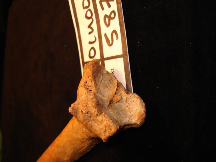
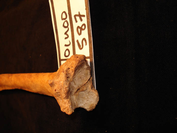
| OCU00
|
587
|
2
|
OCU00_587_2.jpg
|
Healed Colles fracture of the distal end of the right radius showing joint surface and joint margin changes
|
| OCU00
|
587
|
3
|
OCU00_587_3.jpg
|
Healed Colles fracture of the distal end of the right radius showing joint surface and joint margin changes
|
| OCU00
|
593
|
1
|
OCU00_593_1.jpg
|
Natural depression on visceral surface of ribs
|
| OCU00
|
593
|
2
|
OCU00_593_2.jpg
|
Natural depression on visceral surface of ribs
|
| OCU00
|
593
|
3
|
OCU00_593_3.jpg
|
Healed fracture of distal right radius with secondary infection
|
| OCU00
|
593
|
4
|
OCU00_593_4.jpg
|
Osteoarthritis of distal interphlangeal joint
|
| OCU00
|
593
|
5
|
OCU00_593_5.jpg
|
Osteoarthritis of distal interphlangeal joint
|
| OCU00
|
593
|
6
|
OCU00_593_6.jpg
|
Osteoarthritis of distal interphlangeal joint
|
| OCU00
|
593
|
7
|
OCU00_593_7.jpg
|
Healed fracture of MC1 with secondary osteoarthritis (palmar view)
|
| OCU00
|
593
|
8
|
OCU00_593_8.jpg
|
Healed fracture of MC1 with secondary osteoarthritis (lateral view)
|
| OCU00
|
593
|
9
|
OCU00_593_9.jpg
|
Healed fracture of 1st distal phalange (dorsal view)
|
| OCU00
|
593
|
10
|
OCU00_593_10.jpg
|
Healed fracture of 1st distal phalange (lateral view)
|
| OCU00
|
593
|
11
|
OCU00_593_11.jpg
|
Bilateral circular defects of patellae
|
| OCU00
|
593
|
12
|
OCU00_593_12.jpg
|
Osteoarthritis of left proximal tibia
|
| OCU00
|
593
|
13
|
OCU00_593_13.jpg
|
Frilly changes to distal phalanges of the hand
|
| OCU00
|
593
|
14
|
OCU00_593_14.jpg
|
Atrophy of distal asepct of 1st distal phalange of the hand (dorsal view)
|
| OCU00
|
593
|
15
|
OCU00_593_15.jpg
|
Atrophy of distal asepct of 1st distal phalange of the hand (lateral view)
|
| OCU00
|
593
|
16
|
OCU00_593_16.jpg
|
Distal phalanges of hand (view of distal end)
|
| OCU00
|
593
|
17
|
OCU00_593_17.jpg
|
Distal phalanges of hand (view of distal end)
|
| OCU00
|
593
|
18
|
OCU00_593_18.jpg
|
Frilly changes to distal phalanges of the hand
|
| OCU00
|
615
|
1
|
OCU00_615_1.jpg
|
Osteomalacia fracture line of left scapula
|
| OCU00
|
615
|
2
|
OCU00_615_2.jpg
|
Osteomalacia fracture line of left scapula
|
| OCU00
|
615
|
3
|
OCU00_615_3.jpg
|
Fragments of vertebra (osteomalacia/osteoporosis)
|
| OCU00
|
615
|
4
|
OCU00_615_4.jpg
|
Unhealed rib fractures caused by osteomalacia
|
| OCU00
|
615
|
5
|
OCU00_615_5.jpg
|
Unhealed rib fractures caused by osteomalacia
|
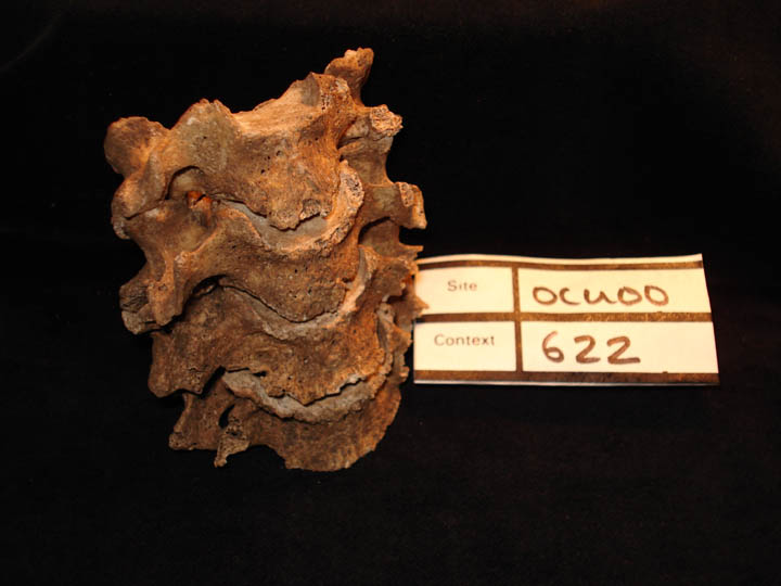
| OCU00
|
622
|
1
|
OCU00_622_1.jpg
|
Cervical vertebrae showing osteophytic lipping (anterior view)
|
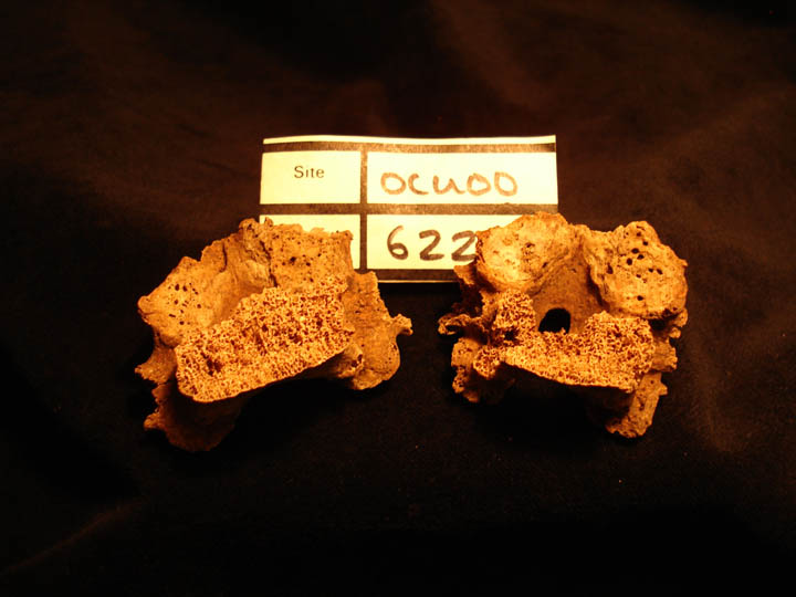
| OCU00
|
622
|
2
|
OCU00_622_2.jpg
|
Cervical vertebrae showing osteophytic lipping,eburnation and destruction of facets (superior & inferior view)
|
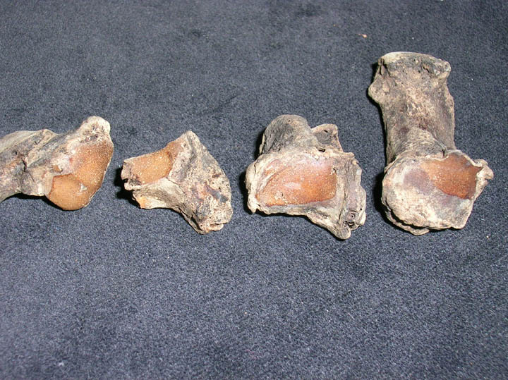
| OCU00
|
622
|
3
|
OCU00_622_3.jpg
|
Thoracic vertebare showing destruction of facets (inferior view)
|
| OCU00
|
654
|
1
|
OCU00_654_1.jpg
|
Osteoarthritis of carpals and metacarpal
|
| OCU00
|
654
|
2
|
OCU00_654_2.jpg
|
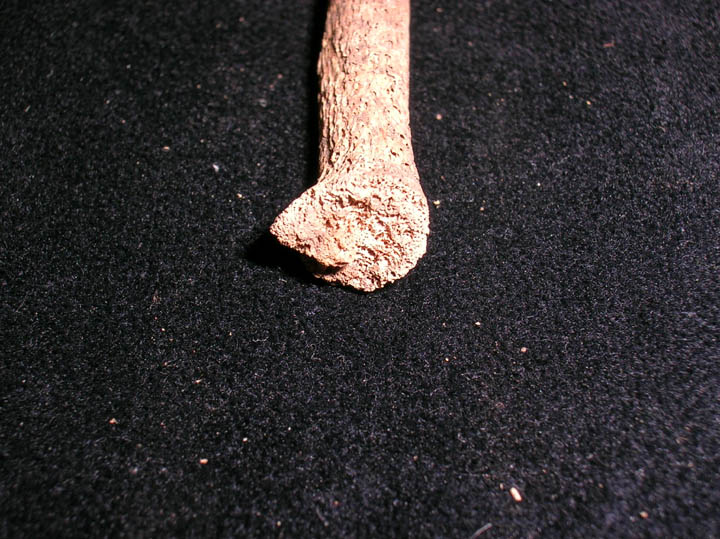
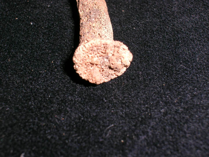
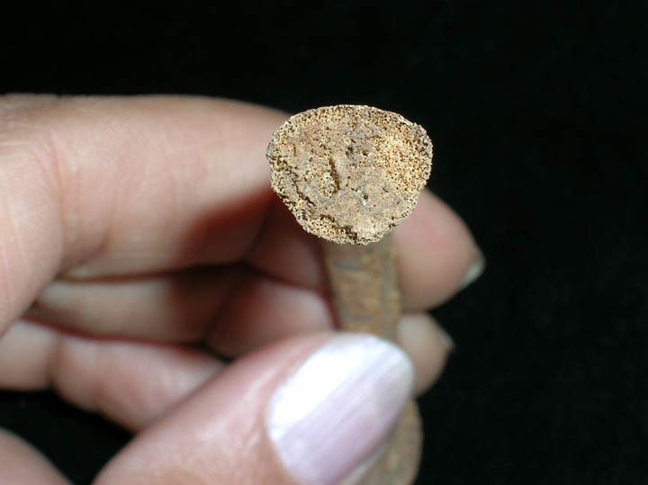
nails
|
| OCU00
|
654
|
3
|
OCU00_654_3.jpg
|
nails
|
| OCU00
|
654
|
4
|
OCU00_654_4.jpg
|
nails
|
| OCU00
|
654
|
5
|
OCU00_654_5.jpg
|
nails
|
| OCU00
|
654
|
6
|
OCU00_654_6.jpg
|
nails
|
| OCU00
|
654
|
7
|
OCU00_654_7.jpg
|
nails
|
| OCU00
|
654
|
8
|
OCU00_654_8.jpg
|
Skull
|
| OCU00
|
654
|
9
|
OCU00_654_9.jpg
|
Left lateral view of skull with nasal fracture and antemortem tooth loss
|
| OCU00
|
654
|
10
|
OCU00_654_10.jpg
|
Anterior view of skull with nasal fracture
|
| OCU00
|
654
|
11
|
OCU00_654_11.jpg
|
Cyst lesion ? On left maxilla
|
| OCU00
|
654
|
12
|
OCU00_654_12.jpg
|
Ankylosis of left sacroilliac joint (posterior view)
|
| OCU00
|
654
|
13
|
OCU00_654_13.jpg
|
Ankylosis of left sacroilliac joint (anterior view)
|
| OCU00
|
654
|
14
|
OCU00_654_14.jpg
|
Osteoarthritis of metacarpal phalangeal joint
|
| OCU00
|
668
|
1
|
OCU00_668_1.jpg
|
Osteophytic lipping on lumbar vertebrae
|
| OCU00
|
668
|
2
|
OCU00_668_2.jpg
|
Osteophytic lipping on lumbar vertebrae
|
| OCU00
|
668
|
3
|
OCU00_668_3.jpg
|
Osteophytic lipping on lumbar vertebrae
|
| OCU00
|
668
|
4
|
OCU00_668_4.jpg
|
Osteoarthritis of metacarpal phalangeal joint
|
| OCU00
|
668
|
5
|
OCU00_668_5.jpg
|
Lytic lesions on the lateral aspect of the MC
|
| OCU00
|
668
|
6
|
OCU00_668_6.jpg
|
Lytic lesions on the lateral aspect of the MC
|
| OCU00
|
668
|
7
|
OCU00_668_7.jpg
|
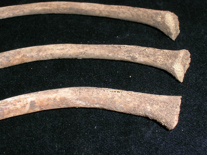
Fibula
|
| OCU00
|
668
|
8
|
OCU00_668_8.jpg
|
Healed fracture of the right illium (posterior view)
|
| OCU00
|
668
|
9
|
OCU00_668_9.jpg
|
Healed fracture of the right illium (anterior view)
|
| OCU00
|
668
|
10
|
OCU00_668_10.jpg
|
Healed fracture of radius
|
| OCU00
|
668
|
11
|
OCU00_668_11.jpg
|
Osteophyte on joint of right humerus (anterior view)
|
| OCU00
|
668
|
12
|
OCU00_668_12.jpg
|
Osteophyte on joint of right humerus (posterior view)
|
| OCU00
|
668
|
13
|
OCU00_668_13.jpg
|
Ribs with costal cartilage
|
| OCU00
|
668
|
14
|
OCU00_668_14.jpg
|
Ribs with costal cartilage
|
| OCU00
|
681
|
1
|
OCU00_681_1.jpg
|
Skull of William Wood showing edentulous maxillae and mandible
|
| OCU00
|
681
|
2
|
OCU00_681_2.jpg
|
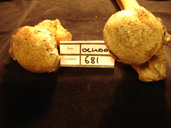
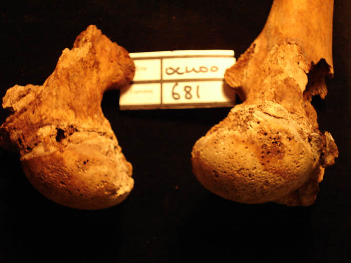
Ox coxae of William Wood showing bilateral osteoarthritis of the acetabulae
|
| OCU00
|
681
|
3
|
OCU00_681_3.jpg
|
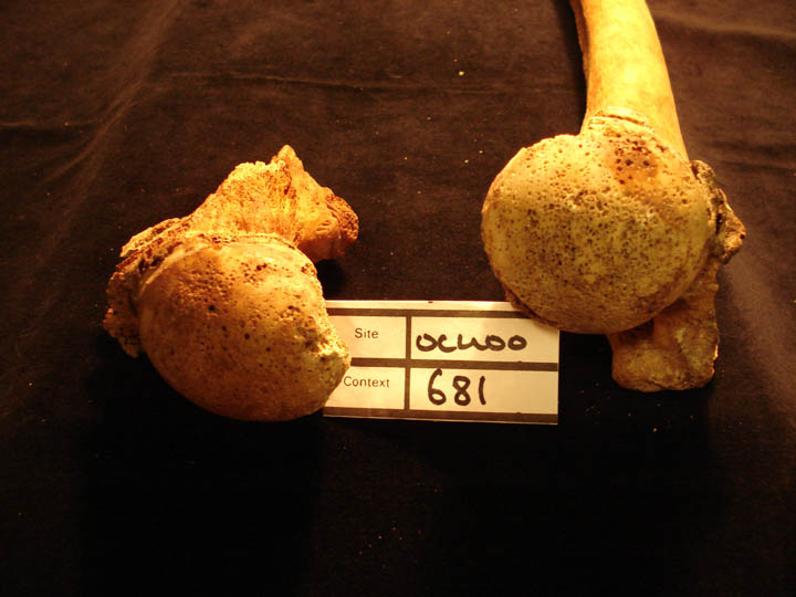
Femora of William Wood showing joint destruction and eburnation of the femoral heads
|
| OCU00
|
681
|
4
|
OCU00_681_4.jpg
|
Femora of William Wood showing joint destruction and eburnation of the femoral heads
|
| OCU00
|
681
|
5
|
OCU00_681_5.jpg
|
Femora of William Wood showing joint destruction and eburnation of the femoral heads
|
| OCU00
|
681
|
6
|
OCU00_681_6.jpg
|
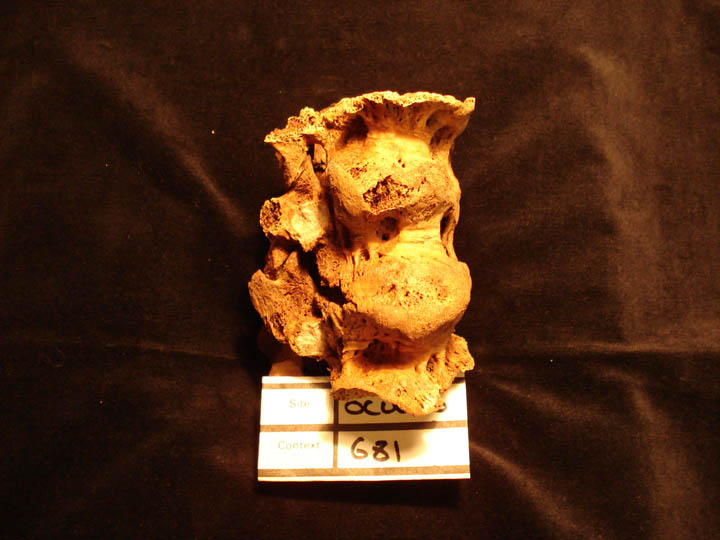
Thoracic vertebare of William Wood showing the classic 'candlewax' fusion of Diffuse Idiopathic Skeletal Hyperostosis (anterior view)
|
| OCU00
|
681
|
7
|
OCU00_681_7.jpg
|
Thoracic vertebrae of William Wood showing the classic 'candlewax' fusion of Diffuse Idiopathic Skeletal Hyperostosis (right side)
|
| OCU00
|
697
|
1
|
OCU00_697_1.jpg
|
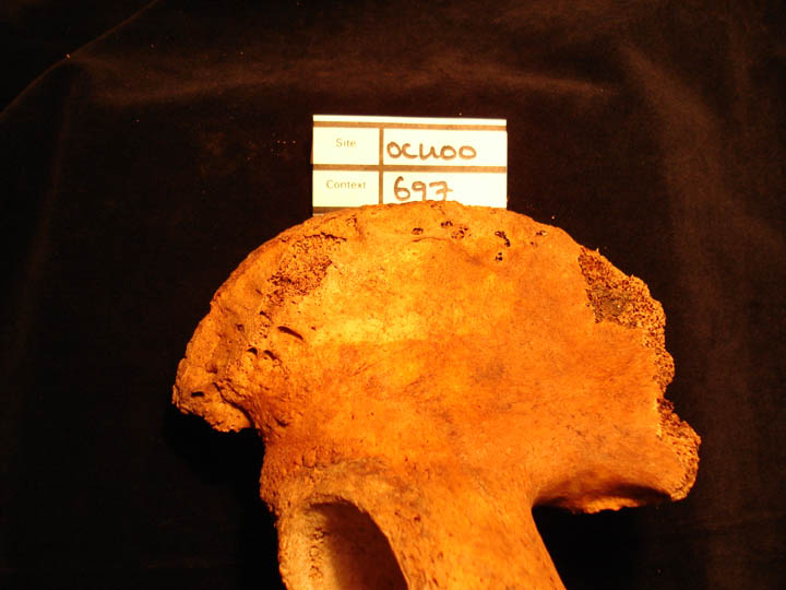
Healed fracture of right pelvis of the ilium (posterior view)
|
| OCU00
|
697
|
2
|
OCU00_697_2.jpg
|
Healed fracture of the right pelvis of the ilium (posterior view)
|
| OCU00
|
697
|
3
|
OCU00_697_3.jpg
|
Humerii showing asymmetry
|
| OCU00
|
701
|
1
|
OCU00_701_1.jpg
|
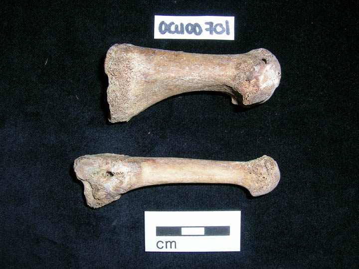
Round lesions on the lateral aspect of MCPHJ of right metatarsals
|
| OCU00
|
701
|
2
|
OCU00_701_2.jpg
|
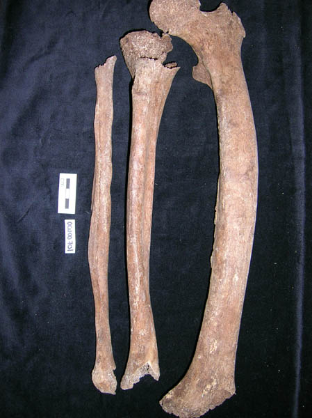
Residual rickets seen in left leg bones
|
| OCU00
|
701
|
3
|
OCU00_701_3.jpg
|
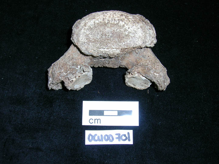
Congenital defect of second cervical vertebra
|
| OCU00
|
701
|
4
|
OCU00_701_4.jpg
|
Spondylolisis of 6th Lumbar vertebra
|
| OCU00
|
701
|
5
|
OCU00_701_5.jpg
|
Possible osteomalacia causing collapse of the sacrum
|
| OCU00
|
701
|
6
|
OCU00_701_6.jpg
|
Bilateral ankylosis of sacroilliac joint
|
| OCU00
|
701
|
7
|
OCU00_701_7.jpg
|
Superior view of skull with hair
|
| OCU00
|
701
|
8
|
OCU00_701_8.jpg
|
Superior view of skull with hair
|
| OCU00
|
701
|
9
|
OCU00_701_9.jpg
|
Base of skull
|
| OCU00
|
701
|
10
|
OCU00_701_10.jpg
|
Base of skull
|
| OCU00
|
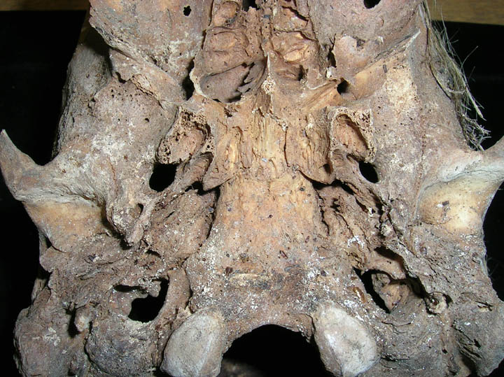
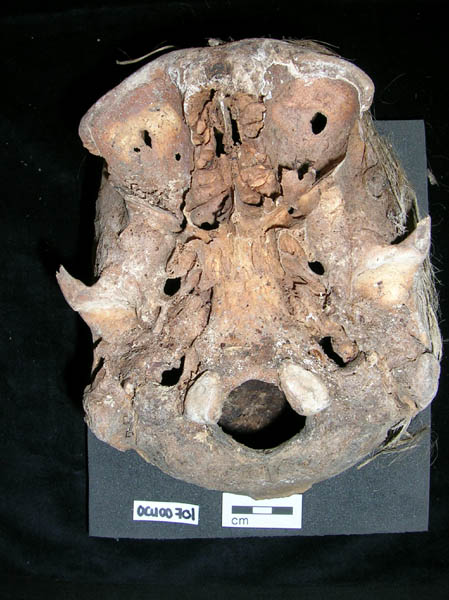
713
|
1
|
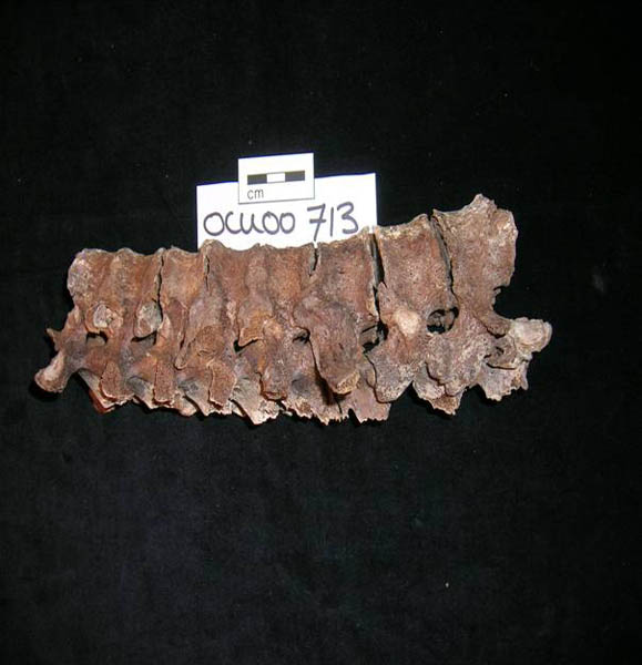
OCU00_713_1.jpg
|
Ankylosing Spondylolylitis of vertebral column (posterior view) John Long Esq
|
| OCU00
|
713
|
2
|
OCU00_713_2.jpg
|
Ankylosing Spondylolylitis of vertebral column (right side view) John Long Esq
|
| OCU00
|
713
|
3
|
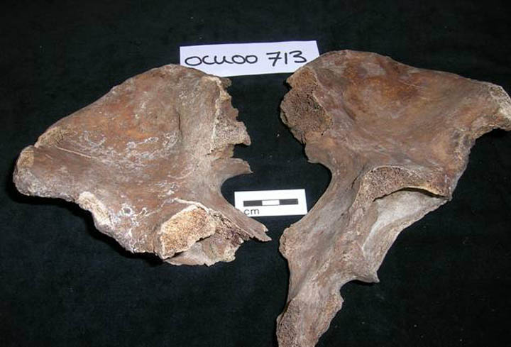
OCU00_713_3.jpg
|
Os Coxae indicating bilateral ankylosis of the sacroiliac joints John Long Esq
|
| OCU00
|
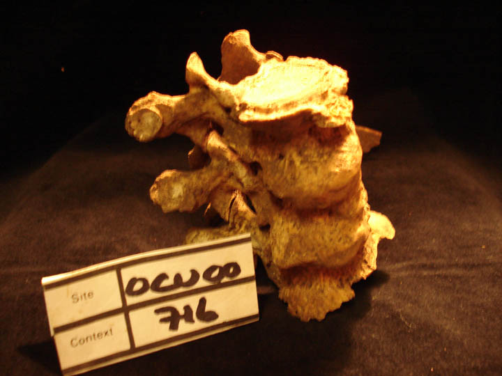
716
|
1
|
OCU00_716_1.jpg
|
Thoracic vertebrae showing the classic 'candlewax' fusion of Diffuse Idiopathic Skeletal Hyperostosis (anterior view)
|
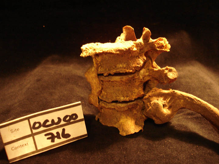
| OCU00
|
716
|
2
|
OCU00_716_2.jpg
|
Thoracic vertebrae showing the classic 'candlewax' fusion of Diffuse Idiopathic Skeletal Hyperostosis and costovertebral fusion on the left side (left side view)
|
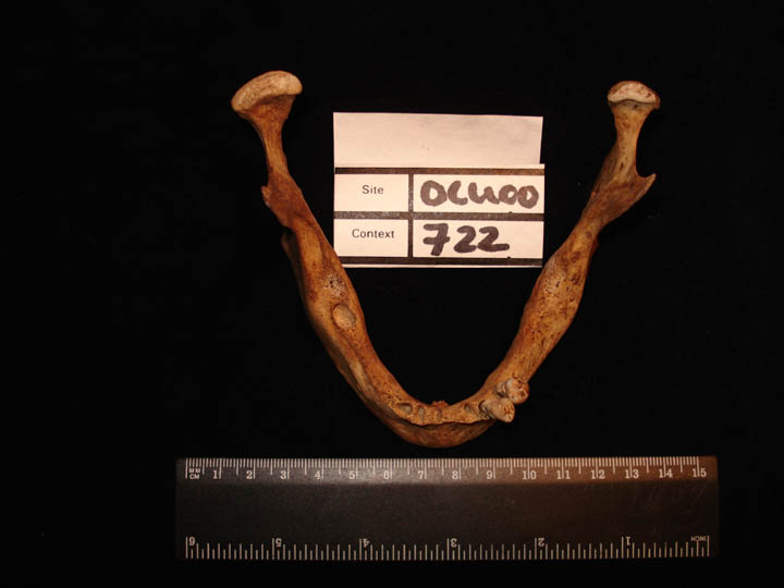
| OCU00
|
722
|
1
|
OCU00_722_1.jpg
|
Reduction and diminished size of the left ramus head
|
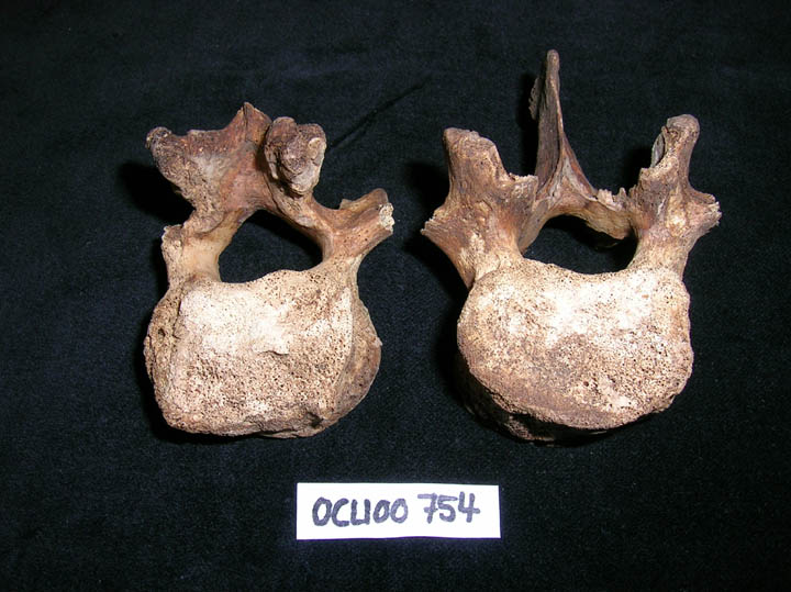
| OCU00
|
754
|
1
|
OCU00_754_1.jpg
|
Tuberculosis changes on L2-3
|
| OCU00
|
764
|
1
|
OCU00_764_1.jpg
|
Osteomalacia fracture line of right scapula
|
| OCU00
|
764
|
2
|
OCU00_764_2.jpg
|
Osteomalacia fracture line of right scapula
|
| OCU00
|
764
|
3
|
OCU00_764_3.jpg
|
Osteomalacia fracture line of right scapula
|
| OCU00
|
764
|
4
|
OCU00_764_4.jpg
|
Osteomalacia fracture of rib
|
| OCU00
|
764
|
5
|
OCU00_764_5.jpg
|
Osteomalacia fracture of rib
|
| OCU00
|
764
|
6
|
OCU00_764_6.jpg
|
Osteomalacia fracture of rib
|
| OCU00
|
764
|
7
|
OCU00_764_7.jpg
|
Endocranial porosity
|
| OCU00
|
764
|
8
|
OCU00_764_8.jpg
|
Endocranial porosity
|
| OCU00
|
764
|
9
|
OCU00_764_9.jpg
|
Untitled
|
| OCU00
|
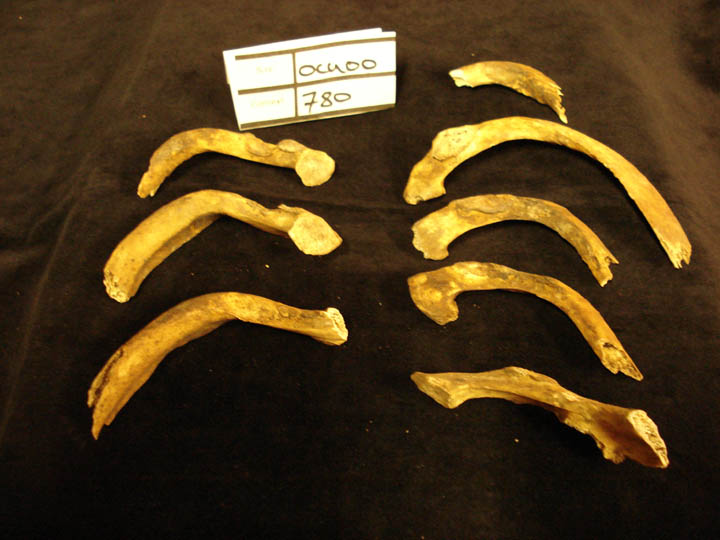
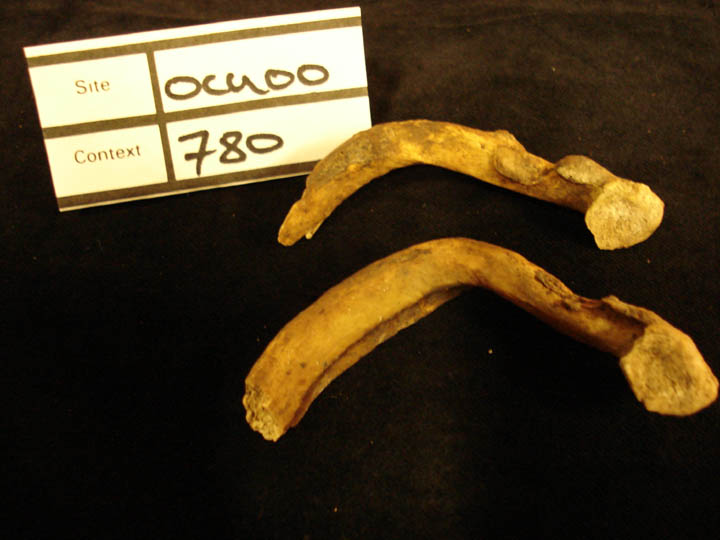
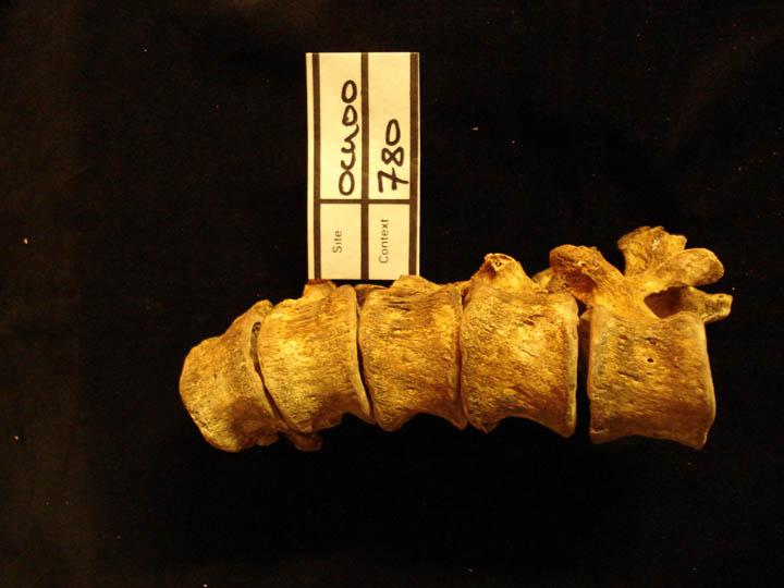
780
|
1
|
OCU00_780_1.jpg
|
Deformity and curvature of ribs L & R from kyphoscoliosis
|
| OCU00
|
780
|
2
|
OCU00_780_2.jpg
|
Deformity and curvature of ribs L & R from kyphoscoliosis
|
| OCU00
|
780
|
3
|
OCU00_780_3.jpg
|
Deformity and curvature of ribs L & R from kyphoscoliosis
|
| OCU00
|
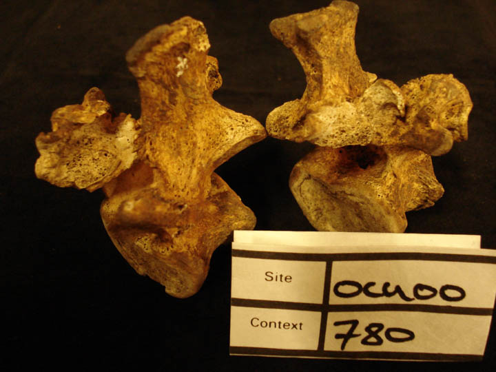
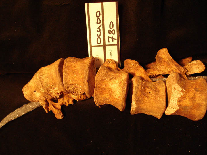
780
|
4
|
OCU00_780_4.jpg
|
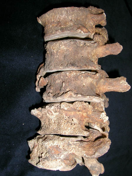
Thoracic vertebrae showing distortion of the articular facets caused by kyphoscoliosis
|
| OCU00
|
780
|
5
|
OCU00_780_5.jpg
|
Thoracic vertebrae showing lateral wedging of the centrums kyphoscoliosis
|
| OCU00
|
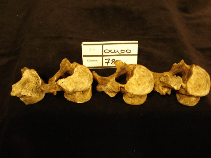
780
|
6
|
OCU00_780_6.jpg
|
Articulated thoracic vertebrae showing curvature of the spine (anterior view) kyphoscoliosis
|
| OCU00
|
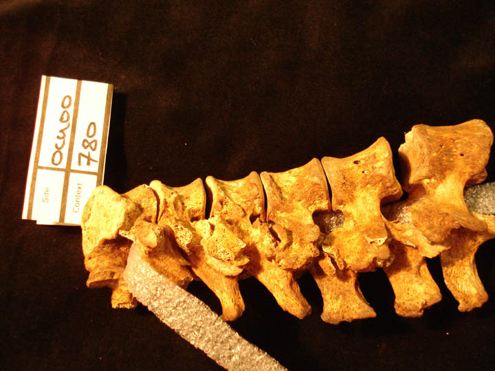
780
|
7
|
OCU00_780_7.jpg
|
Articulated thoracic vertebrae showing curvature of the spine (anterior view) kyphoscoliosis
|
| OCU00
|
780
|
8
|
OCU00_780_8.jpg
|
Articulated thoracic vertebrae showing curvature of the spine (anterior view) kyphoscoliosis
|
| OCU00
|
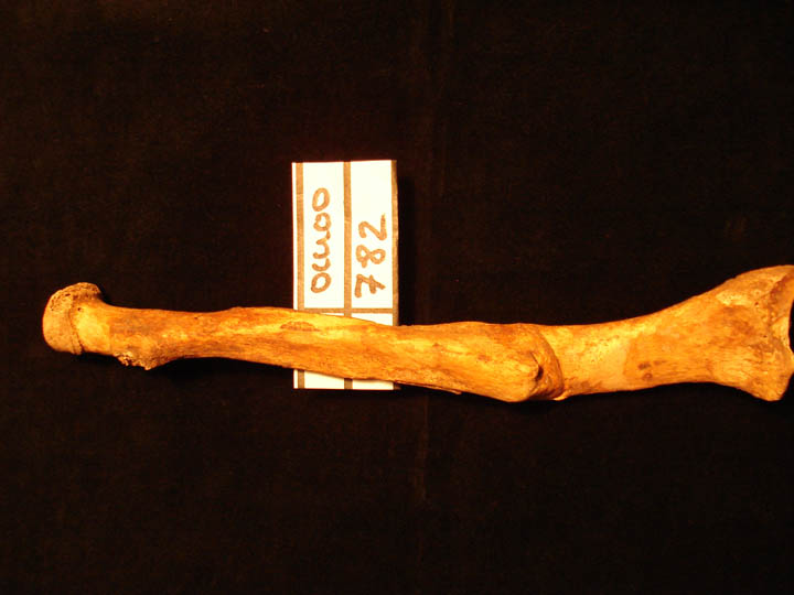
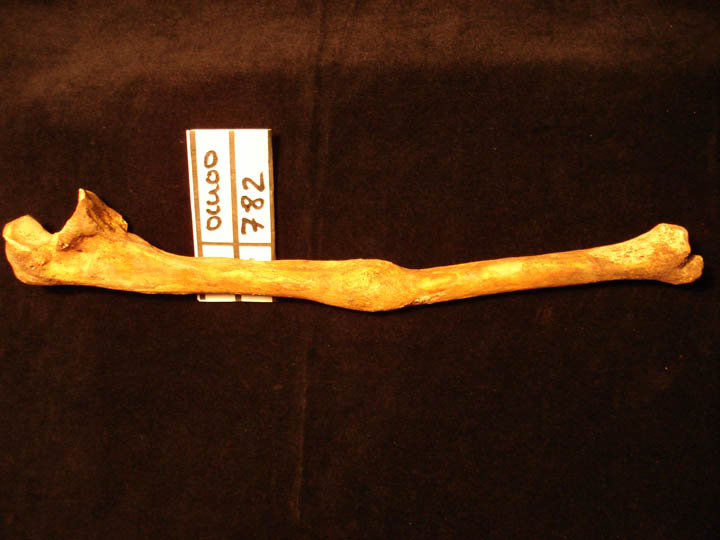
782
|
1
|
OCU00_782_1.jpg
|
Healed fracture of the left radius (mid to distal 1/3 of shaft) showing overlap (posterior view)
|
| OCU00
|
782
|
2
|
OCU00_782_2.jpg
|
Healed fracture of the left radius (mid to distal 1/3 of shaft) showing overlap (posterior view)
|
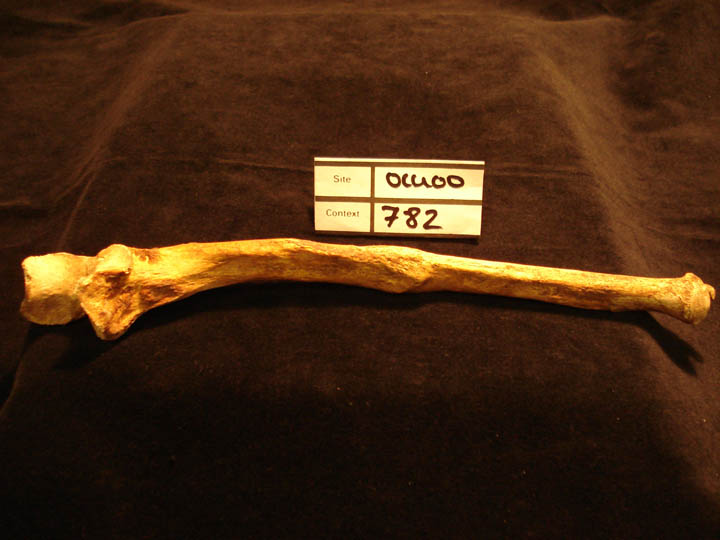
| OCU00
|
782
|
3
|
OCU00_782_3.jpg
|
Healed fracture of the left radius (mid to distal 1/3 of shaft) showing overlap (medial view)
|
| OCU00
|
782
|
4
|
OCU00_782_4.jpg
|
Healed fracture of the left ulna (mid shaft) showing remodelled callus (medial view)
|
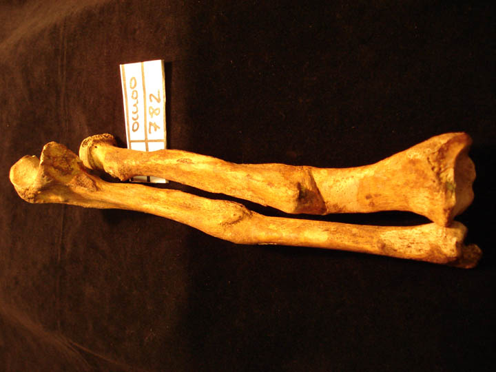
| OCU00
|
782
|
5
|
OCU00_782_5.jpg
|
Healed fracture of the left ulna (mid shaft) showing remodelled callus (anterior view)
|
| OCU00
|
782
|
6
|
OCU00_782_6.jpg
|
Healed fractures of the left ulna and radius articulated (anterior view)
|
| OCU00
|
782
|
7
|
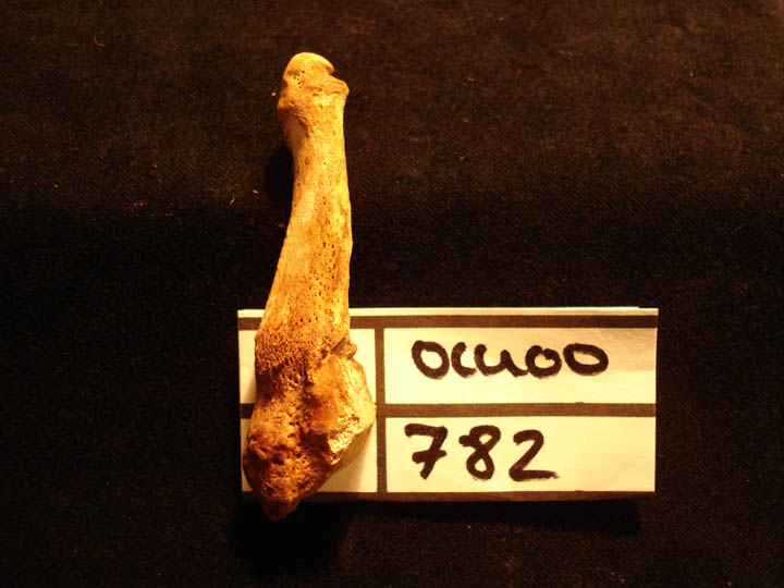
OCU00_782_7.jpg
|
Healed fracture of the left 5th metatarsal distal 1/3 of shaft (anterior view)
|
| OCU00
|
782
|
8
|
OCU00_782_8.jpg
|
Healed fracture of the 5th metatarsal distal 1/3 of shaft (plantar view)
|
| OCU00
|
782
|
9
|
OCU00_782_9.jpg
|
Healed fracture of the 5th metatarsal distal 1/3 of shaft (medial view)
|
| OCU00
|
782
|
10
|
OCU00_782_10.jpg
|
Fracture of lumbar vertebra L4 of the arch possibly unilateral spondylolysis
|
| OCU00
|
782
|
11
|
OCU00_782_11.jpg
|
Thoracic vertebrae spinous processes with healed fractures
|
| OCU00
|
782
|
12
|
OCU00_782_12.jpg
|
Thoracic vertebrae spinous processes (articulated) with healed fractures
|
| OCU00
|
790
|
1
|
OCU00_790_1.jpg
|
perisoteal infection of the visceral surface of the ribs
|
| OCU00
|
790
|
2
|
OCU00_790_2.jpg
|
Erosive arthropathry of left MT1
|
| OCU00
|
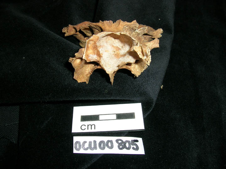
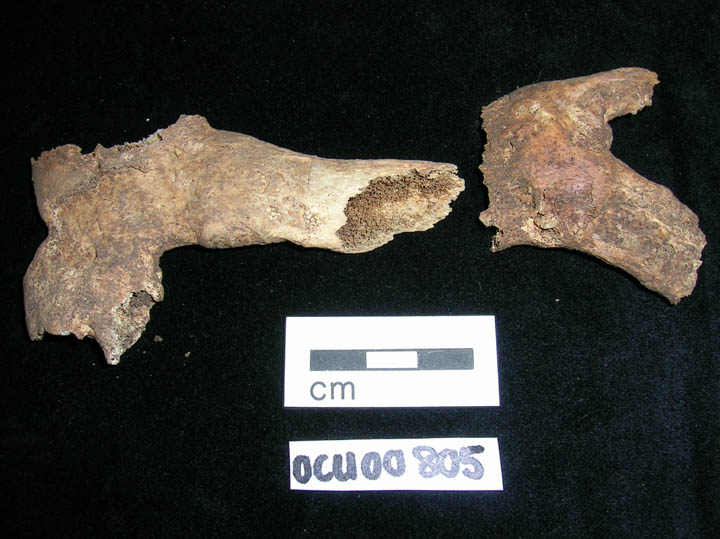
805
|
1
|
OCU00_805_1.jpg
|
Sphenoidal infection
|
| OCU00
|
805
|
2
|
OCU00_805_2.jpg
|
Autopsy calvarium cut on frontal bone
|
| OCU00
|
805
|
3
|
OCU00_805_3.jpg
|
Autopsy calvarium cut on left temporal bone
|
| OCU00
|
805
|
4
|
OCU00_805_4.jpg
|
Autopsy calvarium cut of frontal bone
|
| OCU00
|
805
|
5
|
OCU00_805_5.jpg
|
Costal cartilage
|
| OCU00
|
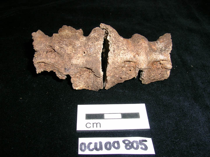
805
|
6
|
OCU00_805_6.jpg
|
DISH
|
| OCU00
|
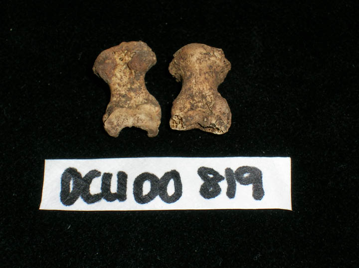
819
|
1
|
OCU00_819_1.jpg
|
Half circular lesions on distal phalanges
|
| OCU00
|
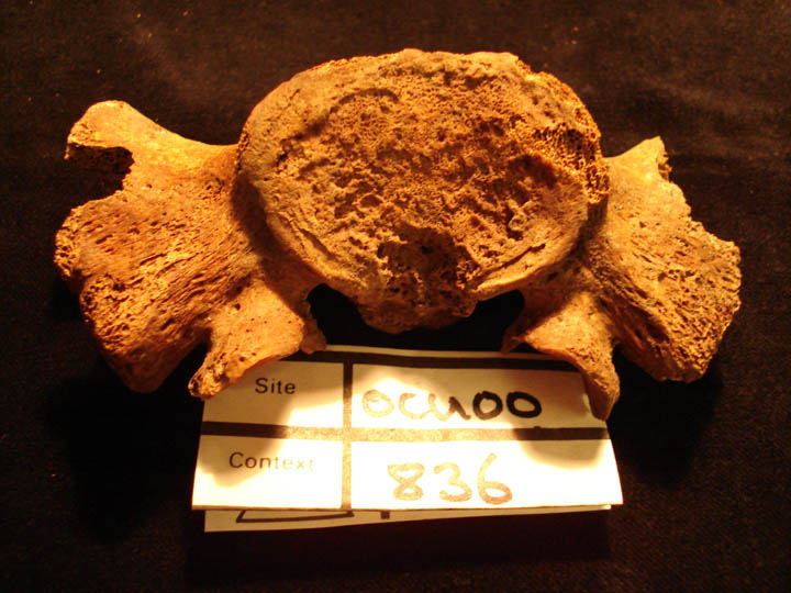
836
|
1
|
OCU00_836_1.jpg
|
Possible lytic lesion (?TB) of the superior body of the 1st sacral vertebra (posterior view)
|
| OCU00
|
836
|
2
|
OCU00_836_2.jpg
|
Possible lytic lesion (?TB) of the superior body of the 1st sacral vertebra (superior view)
|
| OCU00
|
836
|
3
|
OCU00_836_3.jpg
|
Possible lytic lesion (?TB) of the superior body of the 1st sacral vertebra (superior view)
|
| OCU00
|
841
|
1
|
OCU00_841_1.jpg
|
Hyperostosis frontalis interna (early stage)
|
| OCU00
|
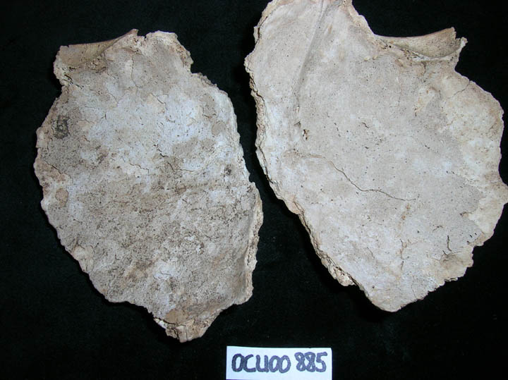
885
|
1
|
OCU00_885_1.jpg
|
Meningeal infection
|
| OCU00
|
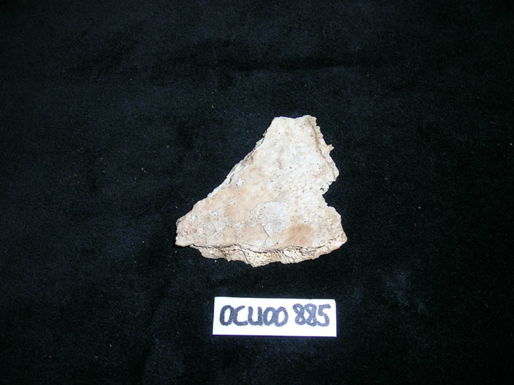
885
|
2
|
OCU00_885_2.jpg
|
Meningeal infection
|
| OCU00
|
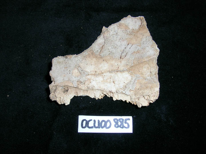
885
|
3
|
OCU00_885_3.jpg
|
Meningeal infection
|
| OCU00
|
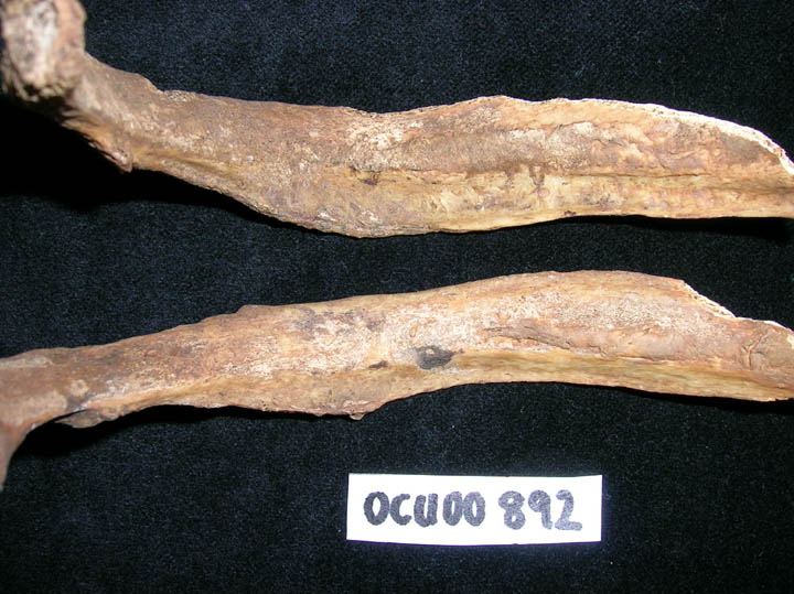
892
|
1
|
OCU00_892_1.jpg
|
Periosteal new bone on visceral surface of ribs
|
| OCU00
|
898
|
1
|
OCU00_898_1.jpg
|
Osteoarthritis of first CMC joint
|
| OCU00
|
898
|
2
|
OCU00_898_2.jpg
|
Bilateral ankylosis sacroilliac joints
|
| OCU00
|
898
|
3
|
OCU00_898_3.jpg
|
Myositis ossificans on posterior proximal portion of left tibia
|
| OCU00
|
898
|
4
|
OCU00_898_4.jpg
|
Bone formation on tarsal bones
|
| OCU00
|
910
|
1
|
OCU00_910_1.jpg
|
Osteoarthrits of the lumbar sacral joint facets
|
| OCU00
|
910
|
2
|
OCU00_910_2.jpg
|
Residual rickets seen in left femur
|
| OCU00
|
910
|
3
|
OCU00_910_3.jpg
|
Residual rickets seen in left and right femora
|
| OCU00
|
910
|
4
|
OCU00_910_4.jpg
|
Residual rickets seen in left and right femora
|
| OCU00
|
910
|
5
|
OCU00_910_5.jpg
|
Residual rickets seen in left and right fibulae
|
| OCU00
|
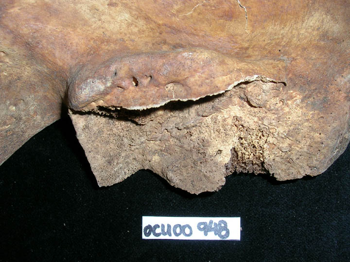
948
|
1
|
OCU00_948_1.jpg
|
Healed fracture of left MT2
|
| OCU00
|
948
|
2
|
OCU00_948_2.jpg
|
Healed fracture of right humeral head
|
| OCU00
|
948
|
3
|
OCU00_948_3.jpg
|
Healed fracture of right humeral head
|
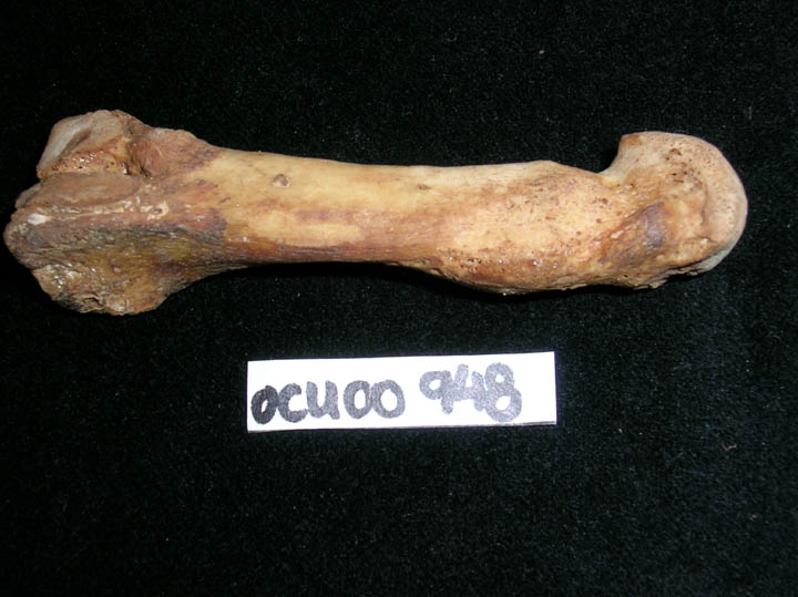
| OCU00
|
948
|
4
|
OCU00_948_4.jpg
|
Healed fracture of right humeral head
|
| OCU00
|
948
|
5
|
OCU00_948_5.jpg
|
Healed fracture of MC1
|
| OCU00
|
948
|
6
|
OCU00_948_6.jpg
|
Periosteal infection on distal joint of left radius
|
| OCU00
|
948
|
7
|
OCU00_948_7.jpg
|
Hairline fracture of capitate
|
| OCU00
|
948
|
8
|
OCU00_948_8.jpg
|
Hairline fracture of capitate
|
| OCU00
|
948
|
9
|
OCU00_948_9.jpg
|
Ankylosis of right sacroilliac joint
|
| OCU00
|
948
|
10
|
OCU00_948_10.jpg
|
Myositis ossificans on right distal femur
|
| OCU00
|
948
|
11
|
OCU00_948_11.jpg
|
Healed fracture of left MT2
|
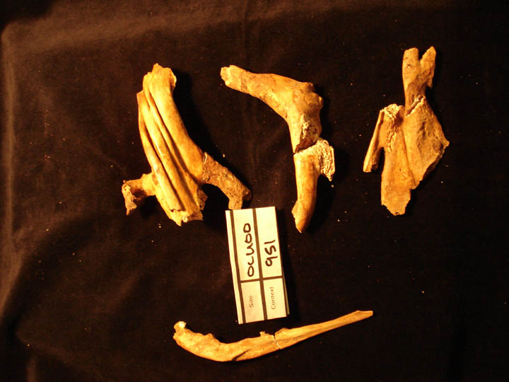
| OCU00
|
951
|
1
|
OCU00_951_1.jpg
|
Gross congenital deformity of left scapula, clavicle and ribs with compression, fusion and distortion
|
| OCU00
|
951
|
2
|
OCU00_951_2.jpg
|
Gross congenital deformity of left scapula, clavicle and ribs with compression, fusion and distortion
|
| OCU00
|
970
|
1
|
OCU00_970_1.jpg
|
Sub adult occipital bone endocranial surface showing localised increase in porosity
|
| OCU00
|
970
|
2
|
OCU00_970_2.jpg
|
Sub adult occipital bone endocranial surface showing localised increase in porosity
|
| OCU00
|
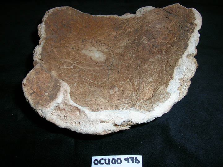
976
|
1
|
OCU00_976_1.jpg
|
Pagets disease in skull
|
| OCU00
|
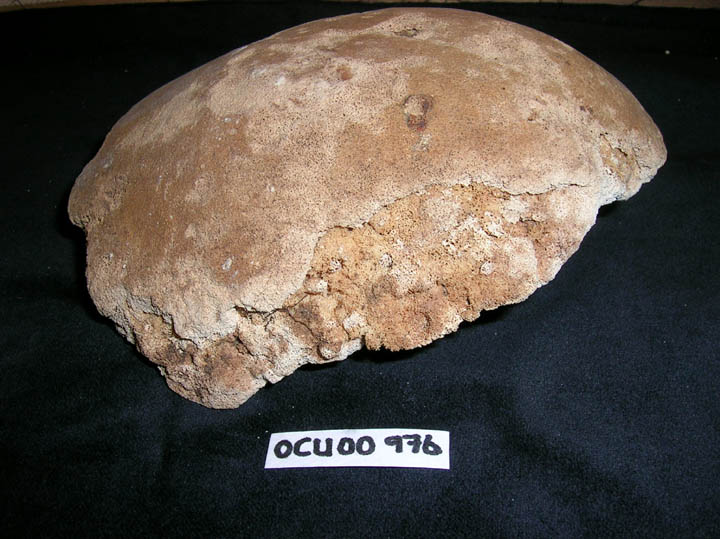
976
|
2
|
OCU00_976_2.jpg
|
Pagets disease in skull
|
| OCU00
|
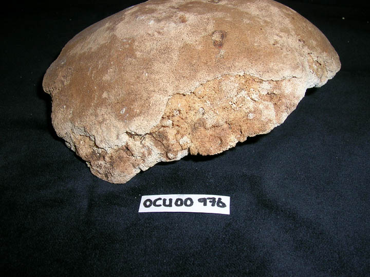
976
|
3
|
OCU00_976_3.jpg
|
Pagets disease in skull
|
| OCU00
|
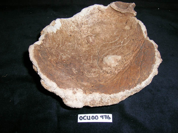
976
|
4
|
OCU00_976_4.jpg
|
Pagets disease in skull
|
| OCU00
|
976
|
5
|
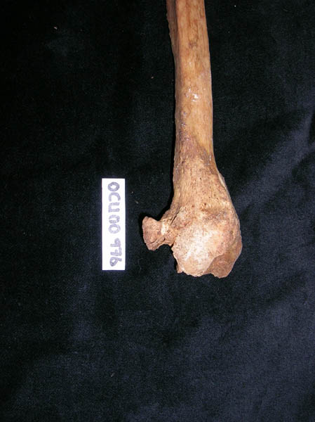
OCU00_976_5.jpg
|
Healed fracture of distal right fibula
|
| OCU00
|
976
|
6
|
OCU00_976_6.jpg
|
Healed fracture of distal right fibula
|
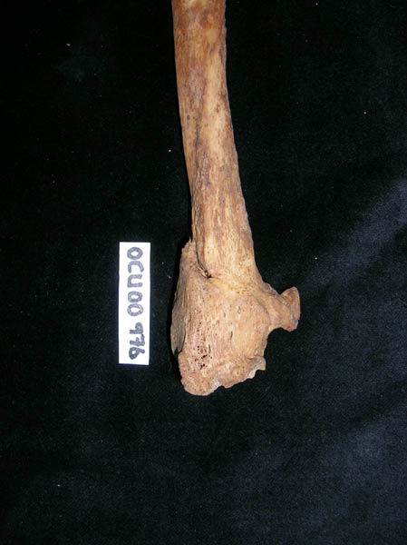
| OCU00
|
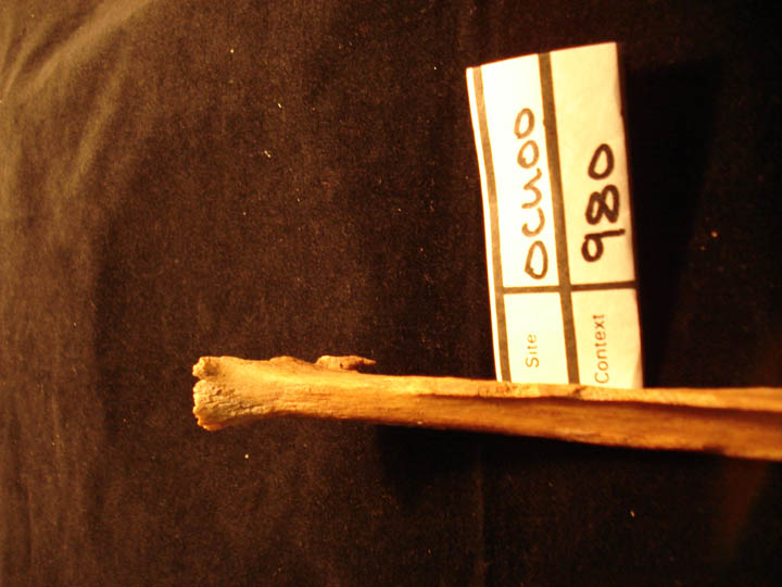
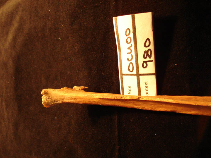
980
|
1
|
OCU00_980_1.jpg
|
Soft tissue trauma myostosis ossificans proximal end of the right fibula
|
| OCU00
|
980
|
2
|
OCU00_980_2.jpg
|
Soft tissue trauma myostosis ossificans proximal end of the right fibula
|
| OCU00
|
1021
|
1
|
OCU00_1021_1.jpg
|
Deformation of sacrum
|
| OCU00
|
1021
|
2
|
OCU00_1021_2.jpg
|
Ankylsosis of metacarpals
|
| OCU00
|
1021
|
3
|
OCU00_1021_3.jpg
|
DISH (right lateral view)
|
| OCU00
|
1021
|
4
|
OCU00_1021_4.jpg
|
Avulsion injury of vertebra
|
| OCU00
|
1021
|
5
|
OCU00_1021_5.jpg
|
Bilateral ankylosis sacroilliac joints
|
| OCU00
|
1021
|
6
|
OCU00_1021_6.jpg
|
Periosteal infection of the visceral surface of the ribs
|
| OCU00
|
1021
|
7
|
OCU00_1021_7.jpg
|
Osteoarthritis of first CMC joint
|
| OCU00
|
1023
|
1
|
OCU00_1023_1.jpg
|
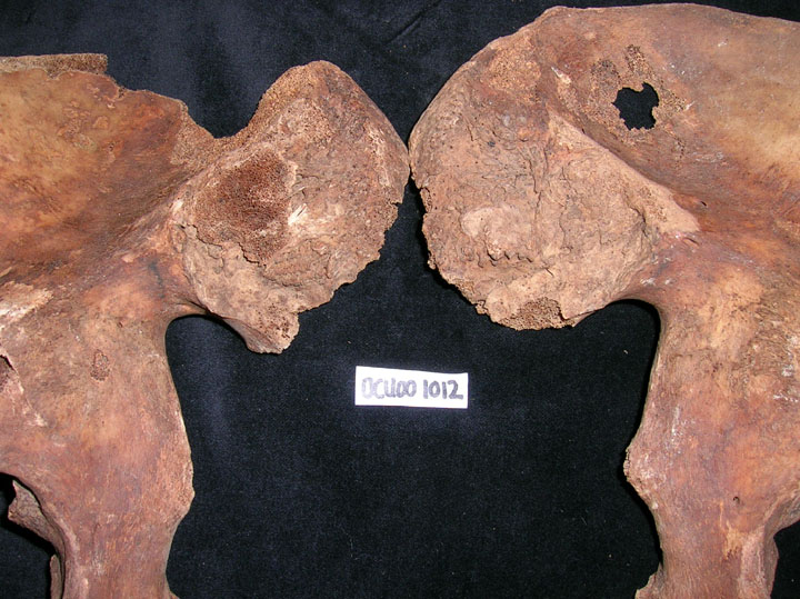
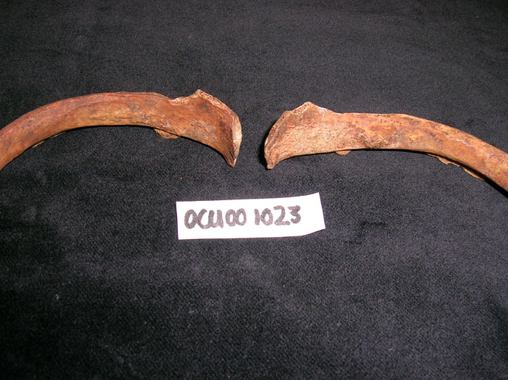
Periosteal infection of the visceral surface of the ribs
|
| OCU00
|
1068
|
1
|
OCU00_1068_1.jpg
|
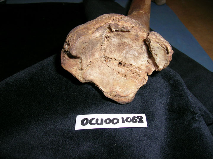
Interarticular fracture left talocural joint
|


